若您需要咨询产品或有任何技术问题,请通过官方电话 400 885 9050 或邮箱 info.cn@stemcell.com 与我们联系。

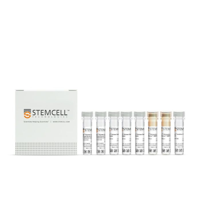
EasySep™ Release 人PSC来源神经嵴细胞正选试剂盒

采用磁珠解离技术,对来源于人多能干细胞分化的神经嵴细胞中的 CD271⁺ 细胞进行免疫磁珠正选分离

只有 %1
¥9,158.00

产品号 #(选择产品)

产品号 #100-0047_C

采用可解离磁珠技术的免疫磁珠正选试剂盒

产品优势

  • 在20分钟内分选出高纯度的人PSC来源神经嵴细胞
  • 无需清洗去除EasySep™ Releasable RapidSpheres™可解离磁珠
  • 和下游分化方案兼容

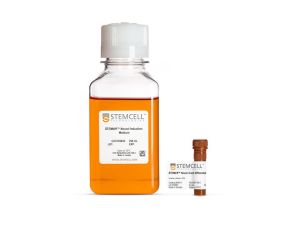
产品组分包括

  • EasySep™ Release人psc衍生的神经嵴细胞正选抗体混合物,2 × 1.0 mL
  • EasySep™分选抗体混合物增强剂,1.0 mL
  • EasySep™ Release缓冲液(浓缩),3 × 1.0 mL
  • EasySep™ Releasable RapidSpheres™ 50201,2 x 1.0 mL
专为您的实验方案打造的产品
要查看实验方案所需的所有配套产品,请参阅《实验方案与技术文档》

总览

使用EasySep™ Release 人PSC来源神经嵴细胞正选试剂盒,通过免疫磁性正选法,可轻松从人多能干细胞(PSC)来源的神经嵴细胞培养样本中分离出高纯度且无磁珠标记的人CD271+细胞。EasySep™技术在已发表的研究中被广泛应用超过20年,将单克隆抗体的特异性与无柱磁珠分选系统的简便性相结合。

在此EasySep™正选步骤中,首先使用可识别CD271的抗体复合物和称为EasySep™ Releasable RapidSpheres™的磁珠对目的细胞进行标记。与传统始终结合在目的细胞上的磁珠不同,该试剂盒中的RapidSpheres™具有可解离特性。通过EasySep™磁极进行分选后,使用解离剂从EasySep™分离的人PSC来源神经嵴细胞中去除结合的磁珠。最终分选获得的细胞组分包含高纯度CD271+细胞,可立即用于下游应用,如流式细胞术或细胞培养。

使用此EasySep™试剂盒分离细胞后,抗体复合物仍结合在细胞表面,可能与Brilliant Violet™抗体偶联物、聚乙二醇修饰蛋白或其他化学相关配体发生相互作用。了解更多关于免疫磁珠EasySep™技术的工作原理。探索更多产品,包括培养基、添加剂、抗体等,为您的实验流程提供优化方案。


磁极兼容性
• EasySep™磁极(产品号 #18000)
 
分类
细胞分选试剂盒
 
细胞类型
神经细胞,PSC衍生
 
种属

 
样本来源
其他组织
 
分选方法
正选
 
应用
细胞分选
 
品牌
EasySep
 
研究领域
疾病建模,神经科学
 

实验数据

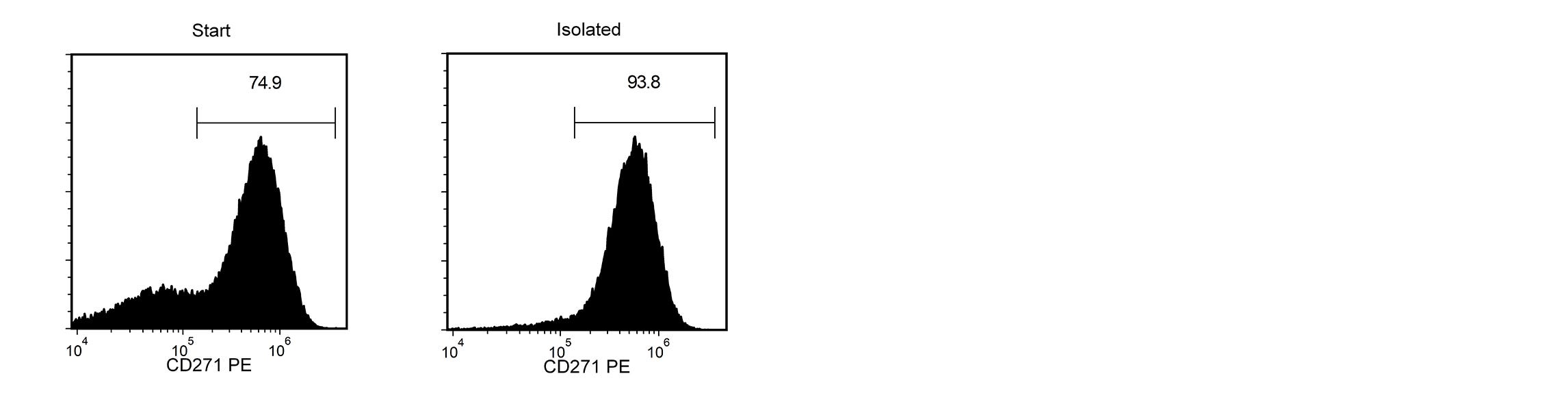
A H9 cell-derived cell suspension assessed for CD271-PE. The the start and final isolated fractions are 74.9% and 93.8% pure.

Figure 1. Histograms of H9 cell suspensions assessed for CD271-phycoerythrin (PE) expression by flow cytometry

Starting with a single-cell suspension of H9 cells differentiated using STEMdiff™ Neural Crest Differentiation Kit (Catalog #08610), the CD271+ cell content of the isolated fractions is typically 92.9 ± 6.3% (mean ± SD using the purple EasySep™ Magnet).

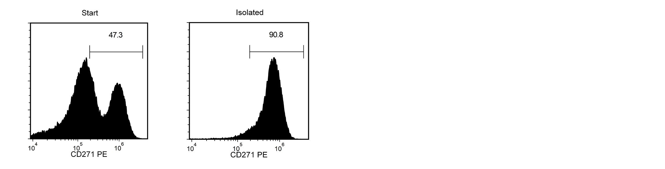
A H7 cell-derived cell suspension assessed for CD271-PE. The the start and final isolated fractions are 47.3% and 90.8% pure.

Figure 2. Histograms of H7 cell suspensions assessed for CD271-phycoerythrin (PE) expression by flow cytometry

Starting with a single-cell suspension of H7 cells differentiated using STEMdiff™ Neural Crest Differentiation Kit (Catalog #08610), the CD271+ cell content of the isolated fractions is typically 92.9 ± 6.3% (mean ± SD using the purple EasySep™ Magnet).

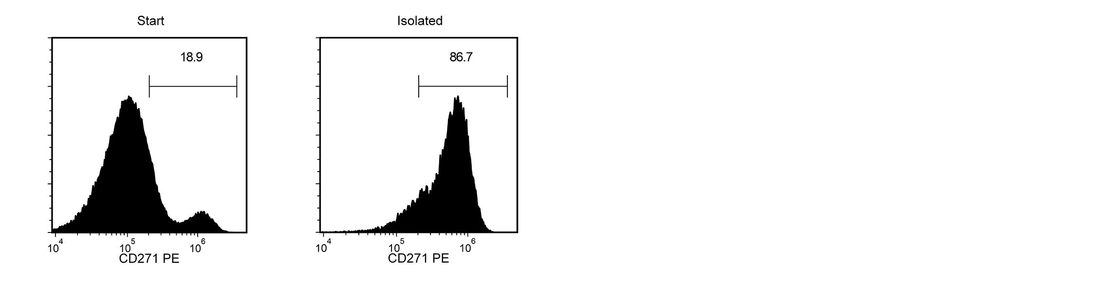
A 1C cell-derived cell suspension assessed for CD271-PE. The the start and final isolated fractions are 18.9% and 86.7% pure.

Figure 3. Histograms of 1C cell suspensions assessed for CD271-phycoerythrin (PE) expression by flow cytometry

Starting with a single-cell suspension of 1C cells differentiated using STEMdiff™ Neural Crest Differentiation Kit (Catalog #08610), the CD271+ cell content of the isolated fractions is typically 92.9 ± 6.3% (mean ± SD using the purple EasySep™ Magnet).

产品说明书及文档

请在《产品说明书》中查找相关支持信息和使用说明,或浏览下方更多实验方案。

Document Type
Product Name
Catalog #
Lot #
Language
Catalog #
100-0047
Lot #
All
Language
English
Document Type
Safety Data Sheet 1
Catalog #
100-0047
Lot #
All
Language
English
Document Type
Safety Data Sheet 2
Catalog #
100-0047
Lot #
All
Language
English
Document Type
Safety Data Sheet 3
Catalog #
100-0047
Lot #
All
Language
English
Document Type
Safety Data Sheet 4
Catalog #
100-0047
Lot #
All
Language
English

应用领域

本产品专为以下研究领域设计,适用于工作流程中的高亮阶段。探索这些工作流程,了解更多我们为各研究领域提供的其他配套产品。

相关材料与文献

技术资料 (9)

文献 (2)

Craniofacial chondrogenesis in organoids from human stem cell-derived neural crest cells iScience 2024 Mar

Abstract

SummaryKnowledge of cell signaling pathways that drive human neural crest differentiation into craniofacial chondrocytes is incomplete,yet essential for using stem cells to regenerate craniomaxillofacial structures. To accelerate translational progress,we developed a differentiation protocol that generated self-organizing craniofacial cartilage organoids from human embryonic stem cell-derived neural crest stem cells. Histological staining of cartilage organoids revealed tissue architecture and staining typical of elastic cartilage. Protein and post-translational modification (PTM) mass spectrometry and snRNA-seq data showed that chondrocyte organoids expressed robust levels of cartilage extracellular matrix (ECM) components: many collagens,aggrecan,perlecan,proteoglycans,and elastic fibers. We identified two populations of chondroprogenitor cells,mesenchyme cells and nascent chondrocytes,and the growth factors involved in paracrine signaling between them. We show that ECM components secreted by chondrocytes not only create a structurally resilient matrix that defines cartilage,but also play a pivotal autocrine cell signaling role in determining chondrocyte fate. Graphical abstract Highlights•Craniofacial cartilage organoids were grown from human neural crest stem cells•These organoids exhibited elastic cartilage architecture and characteristic markers•Paracrine signaling drove chondrogenesis in mesenchyme cells and nascent chondrocytes•ECM components cemented chondrocyte cell fate through autocrine signaling Natural sciences; Biological sciences; Biochemistry; Cell biology; Stem cells research; Specialized functions of cells
Protocol for generating human craniofacial cartilage organoids from stem-cell-derived neural crest cells STAR Protocols 2024 Dec

Abstract

SummaryHere,we present a protocol to generate craniofacial cartilage organoids from human stem cells via neural crest stem cells (NCSCs). We describe steps for inducing human embryonic stem cells (hESCs) or induced pluripotent stem cells (iPSCs) to form NCSCs using sequential treatments of small molecules and growth factors and isolating NCSCs by magnetic bead sorting. We then detail procedures for defining conditions where NCSCs migrate together and self-organize into craniofacial cartilage organoids. Recapitulating craniofacial chondrogenesis will facilitate craniofacial reconstruction and disease modeling.For complete details on the use and execution of this protocol,please refer to Foltz et al.1 Graphical abstract Highlights•Protocol for inducing hESCs or iPSCs to form neural crest stem cells (NCSCs)•Steps for differentiating NCSCs into craniofacial cartilage organoids•Instructions for preparing appropriate media and conditions for differentiation•Guidance for assessing changes in cell and organoid morphology during differentiation Publisher’s note: Undertaking any experimental protocol requires adherence to local institutional guidelines for laboratory safety and ethics. Here,we present a protocol to generate craniofacial cartilage organoids from human stem cells via neural crest stem cells (NCSCs). We describe steps for inducing human embryonic stem cells (hESCs) or induced pluripotent stem cells (iPSCs) to form NCSCs using sequential treatments of small molecules and growth factors and isolating NCSCs by magnetic bead sorting. We then detail procedures for defining conditions where NCSCs migrate together and self-organize into craniofacial cartilage organoids. Recapitulating craniofacial chondrogenesis will facilitate craniofacial reconstruction and disease modeling.

更多信息

更多信息
物种
Magnet Compatibility • EasySep™ Magnet (Catalog #18000)
样本来源 其它细胞系
Selection Method Positive
质量保证:

产品仅供研究使用,不用于针对人或动物的诊断或治疗。 欲获悉更多关于STEMCELL的质控信息,请访问 STEMCELL.CN/COMPLIANCE.
Copyright © 2026 by STEMCELL Technologies. All rights reserved.

在线联系