若您需要咨询产品或有任何技术问题,请通过官方电话 400 885 9050 或邮箱 info.cn@stemcell.com 与我们联系。

EasySep™小鼠T细胞分选试剂盒

通过免疫磁珠负选获得无磁珠标记的小鼠T细胞

只有 %1
¥9,148.00

产品号 #(选择产品)

产品号 #19851_C

通过免疫磁珠负选获得无磁珠标记的小鼠T细胞

产品优势

  •  操作简单、快速
  • 纯度高达99%
  • 无需分离柱
  • 获得不带标记的活细胞

产品组分包括

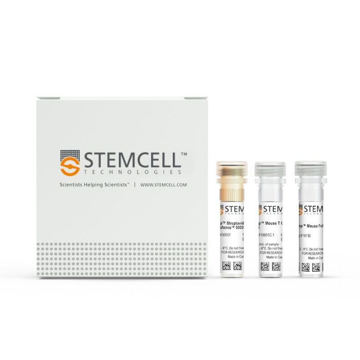
  • EasySep™小鼠T细胞分选试剂盒(产品号 #19851)
    • EasySep™小鼠T细胞分选抗体混合物,0.5 mL
    • EasySep™ Streptavidin RapidSpheres™ 50001磁珠,1 mL
    • EasySep™小鼠FcR阻断剂,0.2 mL
  • RoboSep™小鼠T细胞分选试剂盒(产品号 #19851RF)
    • EasySep™小鼠T细胞分选抗体混合物,0.5 mL
    • EasySep™ Streptavidin RapidSpheres™ 50001磁珠,1 mL
    • EasySep™小鼠FcR阻断剂,0.2 mL
    • RoboSep™ 缓冲液(产品号 #20104)
    • RoboSep™过滤吸头(产品号 #20125)
专为您的实验方案打造的产品
要查看实验方案所需的所有配套产品,请参阅《实验方案与技术文档》

总览

使用EasySep™小鼠T细胞分选试剂盒,通过免疫磁珠负选,可简便高效地从脾细胞或其他组织的单细胞悬液中分离高纯度的小鼠T细胞。EasySep™技术结合单克隆抗体的特异性和无柱磁珠分选系统的简便性,已在发表的研究中广泛应用超过20年。

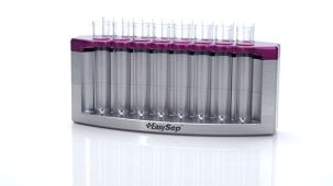
在该EasySep™负选流程中,非目的细胞会被抗体复合物与磁珠标记。表达以下标志物的非目的细胞将被识别并去除:CD11b、CD45R、Ter119、CD49b、CD19和CD24。使用EasySep™磁极吸附后,通过简单地将目的细胞倾倒或吸取至一个新的试管中,即可将被磁珠标记的细胞与不带标记的目的细胞分离开来。在短至15分钟的磁珠分选后,目的T细胞可立即用于流式细胞术、培养及基于细胞的实验等下游应用。

了解更多EasySep™免疫磁珠技术的工作原理,或如何通过RoboSep™实现全自动化免疫磁珠细胞分选。探索为您的实验流程优化的更多产品,包括培养基、添加物、抗体等。


磁极兼容性
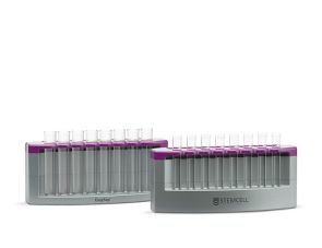
• EasySep™磁极(产品号 #18000)
• “The Big Easy” EasySep™磁极(产品号 #18001)
• EasyEights™ EasySep™磁极(产品号 #18103)
• RoboSep™-S(产品号 #21000)
 
分类
细胞分选试剂盒
 
细胞类型
T 细胞
 
种属
小鼠
 
样本来源
其他组织,脾脏
 
分选方法
负选
 
应用
细胞分选
 
品牌
EasySep,RoboSep
 
研究领域
免疫
 

实验数据

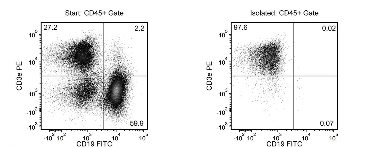
Typical EasySep™ Mouse T Cell Isolation Profile

Figure 1.Typical EasySep™ Mouse T Cell Isolation Profile

Starting with mouse splenocytes, the T cell content (CD3+CD19-) of the isolated fraction is 96.6 ± 2.0% (mean ± SD), using the purple EasySep™ magnet.

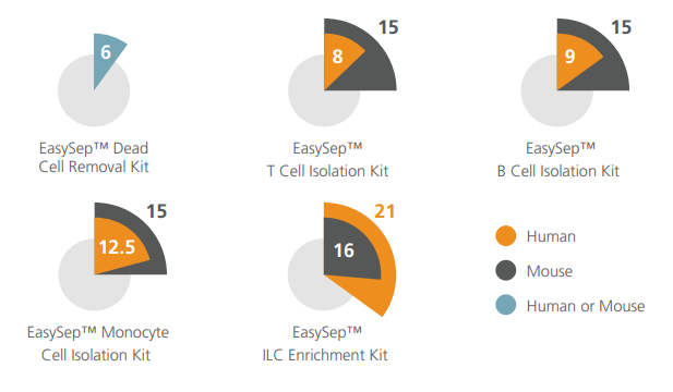
Cell Isolation Protocol Lengths

Figure 2.Cell Isolation Protocol Lengths

Typical time taken (in minutes) to isolate cells using select EasySep™ kits.

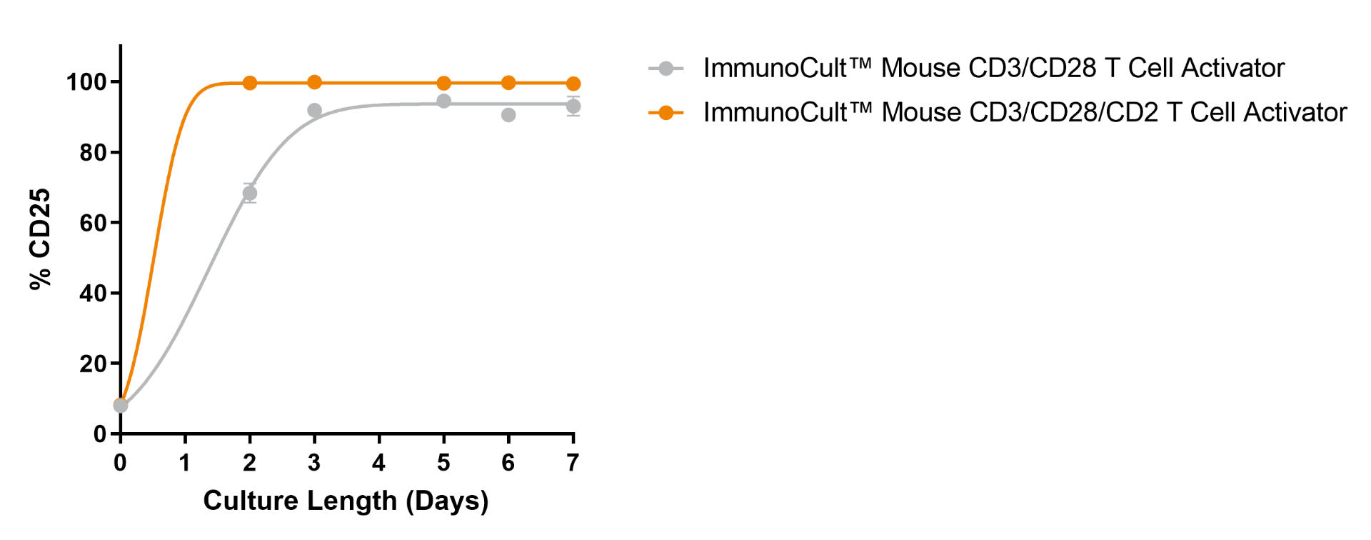
ImmunoCult™ Mouse T Cell Activator Kit Supports High Viability of Activated T Cells

Figure 3.ImmunoCult™ Mouse T Cell Activator Kit Supports High Viability of Activated T Cells

Mouse T cells were isolated using EasySep™ Mouse T Cell Isolation Kit (Catalog #19851), stimulated with ImmunoCult™ Mouse T Cell Activator Kit (Catalog #100-1572), and cultured in IMDM + FBS formulation. Following 3 days of culture, the mean ± SD frequency of CD25+ cells was 91.9 ± 5.1% (n = 11) or 99.9 ± 0.1% (n = 5), when stimulated with ImmunoCult™ Mouse CD3/CD28 T Cell Activator or ImmunoCult™ Mouse CD3/CD28/CD2 T Cell Activator, respectively. Stimulated mouse T cells maintained expression levels of CD25 throughout the 7-day culture period.

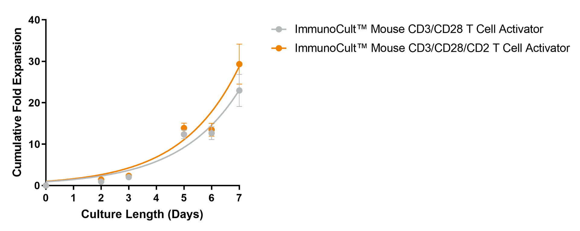
Robust Expansion of EasySep™-Isolated Mouse T Cells Can Be Achieved Following Stimulation with ImmunoCult™ Mouse T Cell Activator Kit

Figure 4.Robust Expansion of EasySep™-Isolated Mouse T Cells Can Be Achieved Following Stimulation with ImmunoCult™ Mouse T Cell Activator Kit

Mouse T cells isolated using EasySep™ Mouse T Cell Isolation Kit (Catalog #19851) were expanded with ImmunoCult™ Mouse T Cell Activator Kit (Catalog #100-1572) in IMDM + FBS formulation over 7 days. The number of viable cells was assessed every 2 - 3 days, and fresh medium supplemented with IL-2 was added. No additional ImmunoCult™ Mouse T Cell Activator was added during the 7-day culture period. After 7 days in culture with ImmunoCult™ Mouse CD3/CD28 T Cell Activator or ImmunoCult™ Mouse CD3/CD28/CD2 T Cell Activator, stimulation resulted in a fold expansion of 23 ± 3.4 or 29.3 ± 4.8 (mean ± SEM, n = 6), respectively.

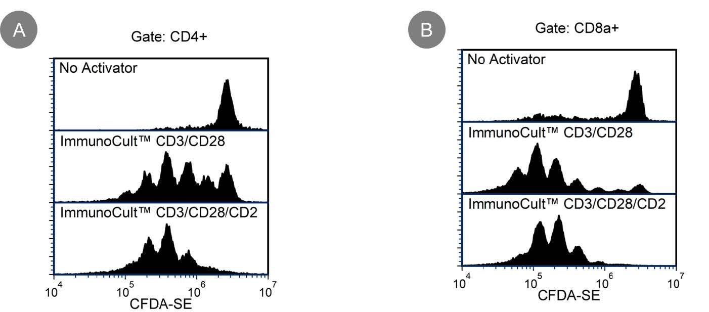
High Cell Proliferation is Observed in EasySep™-Isolated T cells After Stimulation with ImmunoCult™ Mouse T Cell Activator

Figure 5.High Cell Proliferation is Observed in EasySep™-Isolated T cells After Stimulation with ImmunoCult™ Mouse T Cell Activator

Mouse T cells isolated using EasySep™ Mouse T Cell Isolation Kit (Catalog #19851) were labeled with CFDA-SE (Catalog #75003), stimulated with ImmunoCult™ Mouse T Cell Activator Kit (Catalog #100-1572), and cultured in cultured in IMDM + FBS formulation. On Day 3, cells were harvested, stained with anti-mouse CD4 and CD8a antibodies, then measured by flow cytometry. Shown are CFDA-SE-labeled mouse T cells, gated on viable CD4+ (A) or CD8a+ (B) cells, cultured with no activator (top panel), with ImmunoCult™ Mouse CD3/CD28 T Cell Activator (middle panel), or with ImmunoCult™ Mouse CD3/CD28/CD2 T Cell Activator (bottom panel). Due to cell proliferation, the intensity of CFDA-SE signal is reduced by 50% for each cell division.

产品说明书及文档

请在《产品说明书》中查找相关支持信息和使用说明,或浏览下方更多实验方案。

Document Type
Product Name
Catalog #
Lot #
Language
Document Type
产品说明书
Catalog #
19851RF
Lot #
1000120298 or higher
Language
中文
Document Type
产品说明书
Catalog #
19851
Lot #
1000120298 or higher
Language
中文
Catalog #
19851
Lot #
1000120298 or higher
Language
English
Catalog #
19851RF
Lot #
1000120298 or higher
Language
English
Document Type
Safety Data Sheet 1
Catalog #
19851
Lot #
All
Language
English
Document Type
Safety Data Sheet 2
Catalog #
19851
Lot #
All
Language
English
Document Type
Safety Data Sheet 3
Catalog #
19851
Lot #
All
Language
English
Document Type
Safety Data Sheet 4
Catalog #
19851
Lot #
All
Language
English
Document Type
Safety Data Sheet 1
Catalog #
19851RF
Lot #
All
Language
English
Document Type
Safety Data Sheet 2
Catalog #
19851RF
Lot #
All
Language
English
Document Type
Safety Data Sheet 3
Catalog #
19851RF
Lot #
All
Language
English
Document Type
Safety Data Sheet 4
Catalog #
19851RF
Lot #
All
Language
English
Document Type
Safety Data Sheet 5
Catalog #
19851RF
Lot #
All
Language
English

应用领域

本产品专为以下研究领域设计,可应用于工作流程中的关键步骤(图中高亮)。探索这些工作流程,了解更多我们为各研究领域提供的其他配套产品。

相关材料与文献

技术资料 (15)

常见问题 (7)

Can EasySep™ Streptavidin RapidSpheres™ be used for either positive or negative selection?

Currently, EasySep™ Streptavidin RapidSphere™ kits are only available for negative selection and work by targeting and removing unwanted cells.

How does the separation work?

Streptavidin RapidSphere™ magnetic particles are crosslinked to unwanted cells using biotinylated antibodies. When placed in the EasySep™ Magnet, labeled cells migrate to the wall of the tube. The unlabeled cells are then poured off into a new tube.

Which columns do I use?

The EasySep™ procedure is column-free. That's right - no columns!

How can I analyze the purity of my enriched sample?

The Product Information Sheet provided with each EasySep™ kit contains detailed staining information.

Can EasySep™ Streptavidin RapidSphere™ separations be automated?

Yes. RoboSep™, the fully automated cell separator, automates all EasySep™ labeling and cell separation steps.

Are cells isolated using EasySep™ RapidSphere™ products FACS-compatible?

Yes. Desired cells are unlabeled and ready to use in downstream applications, such as FACS analysis.

Can I alter the separation time in the magnet?

Yes; however, this may impact the kit's performance. The provided EasySep™ protocols have already been optimized to balance purity, recovery and time spent on the isolation.

文献 (59)

Deficiency of MALT1 Paracaspase Activity Results in Unbalanced Regulatory and Effector T and B Cell Responses Leading to Multiorgan Inflammation Bornancin F et al. The Journal of Immunology 2015

Abstract

The paracaspase MALT1 plays an important role in immune receptor-driven signaling pathways leading to NF-κB activation. MALT1 promotes signaling by acting as a scaffold,recruiting downstream signaling proteins,as well as by proteolytic cleavage of multiple substrates. However,the relative contributions of these two different activities to T and B cell function are not well understood. To investigate how MALT1 proteolytic activity contributes to overall immune cell regulation,we generated MALT1 protease-deficient mice (Malt1(PD/PD)) and compared their phenotype with that of MALT1 knockout animals (Malt1(-/-)). Malt1(PD/PD) mice displayed defects in multiple cell types including marginal zone B cells,B1 B cells,IL-10-producing B cells,regulatory T cells,and mature T and B cells. In general,immune defects were more pronounced in Malt1(-/-) animals. Both mouse lines showed abrogated B cell responses upon immunization with T-dependent and T-independent Ags. In vitro,inactivation of MALT1 protease activity caused reduced stimulation-induced T cell proliferation,impaired IL-2 and TNF-α production,as well as defective Th17 differentiation. Consequently,Malt1(PD/PD) mice were protected in a Th17-dependent experimental autoimmune encephalomyelitis model. Surprisingly,Malt1(PD/PD) animals developed a multiorgan inflammatory pathology,characterized by Th1 and Th2/0 responses and enhanced IgG1 and IgE levels,which was delayed by wild-type regulatory T cell reconstitution. We therefore propose that the pathology characterizing Malt1(PD/PD) animals arises from an immune imbalance featuring pathogenic Th1- and Th2/0-skewed effector responses and reduced immunosuppressive compartments. These data uncover a previously unappreciated key function of MALT1 protease activity in immune homeostasis and underline its relevance in human health and disease.
CD4 T Cell Help via B Cells Is Required for Lymphopenia-Induced CD8 T Cell Proliferation. Ayasoufi K et al. Journal of Immunology 2016 APR

Abstract

Ab-mediated lymphoablation is commonly used in solid organ and hematopoietic cell transplantation. However,these strategies fail to control pathogenic memory T cells efficiently and to improve long-term transplant outcomes significantly. Understanding the mechanisms of T cell reconstitution is critical for enhancing the efficacy of Ab-mediated depletion in sensitized recipients. Using a murine analog of anti-thymocyte globulin (mATG) in a mouse model of cardiac transplantation,we previously showed that peritransplant lymphocyte depletion induces rapid memory T cell proliferation and only modestly prolongs allograft survival. We now report that T cell repertoire following depletion is dominated by memory CD4 T cells. Additional depletion of these residual CD4 T cells severely impairs the recovery of memory CD8 T cells after mATG treatment. The CD4 T cell help during CD8 T cell recovery depends on the presence of B cells expressing CD40 and intact CD40/CD154 interactions. The requirement for CD4 T cell help is not limited to the use of mATG in heart allograft recipients,and it is observed in nontransplanted mice and after CD8 T cell depletion with mAb instead of mATG. Most importantly,limiting helper signals increases the efficacy of mATG in controlling memory T cell expansion and significantly extends heart allograft survival in sensitized recipients. Our findings uncover the novel role for helper memory CD4 T cells during homeostatic CD8 T cell proliferation and open new avenues for optimizing lymphoablative therapies in allosensitized patients.
Autoantibody-boosted T-cell reactivation in the target organ triggers manifestation of autoimmune CNS disease. Flach A-C et al. Proceedings of the National Academy of Sciences of the United States of America 2016 MAR

Abstract

Multiple sclerosis (MS) is caused by T cells that are reactive for brain antigens. In experimental autoimmune encephalomyelitis,the animal model for MS,myelin-reactive T cells initiate the autoimmune process when entering the nervous tissue and become reactivated upon local encounter of their cognate CNS antigen. Thereby,the strength of the T-cellular reactivation process within the CNS tissue is crucial for the manifestation and the severity of the clinical disease. Recently,B cells were found to participate in the pathogenesis of CNS autoimmunity,with several diverse underlying mechanisms being under discussion. We here report that B cells play an important role in promoting the initiation process of CNS autoimmunity. Myelin-specific antibodies produced by autoreactive B cells after activation in the periphery diffused into the CNS together with the first invading pathogenic T cells. The antibodies accumulated in resident antigen-presenting phagocytes and significantly enhanced the activation of the incoming effector T cells. The ensuing strong blood-brain barrier disruption and immune cell recruitment resulted in rapid manifestation of clinical disease. Therefore,myelin oligodendrocyte glycoprotein (MOG)-specific autoantibodies can initiate disease bouts by cooperating with the autoreactive T cells in helping them to recognize their autoantigen and become efficiently reactivated within the immune-deprived nervous tissue.

更多信息

更多信息
物种 小鼠
Magnet Compatibility • EasySep™ Magnet (Catalog #18000) • “The Big Easy” EasySep™ Magnet (Catalog #18001) • EasyEights™ EasySep™ Magnet (Catalog #18103) • RoboSep™-S (Catalog #21000)
样本来源 其它细胞系, 脾脏
Selection Method Negative
PRODUCTS ARE FOR RESEARCH USE ONLY AND NOT INTENDED FOR HUMAN OR ANIMAL DIAGNOSTIC OR THERAPEUTIC USES UNLESS OTHERWISE STATED. FOR ADDITIONAL INFORMATION ON QUALITY AT STEMCELL, REFER TO WWW.STEMCELL.COM/COMPLIANCE.
Copyright © 2026 by STEMCELL Technologies. All rights reserved.

在线联系