若您需要咨询产品或有任何技术问题,请通过官方电话 400 885 9050 或邮箱 info.cn@stemcell.com 与我们联系。

BrainPhys™ hPSC 神经元试剂盒

用于在BrainPhys™神经元培养基中无血清培养和分化ES/iPS细胞来源的神经元的试剂盒
只有 %1
¥8,510.00

产品号 #(选择产品)

产品号 #05795_C

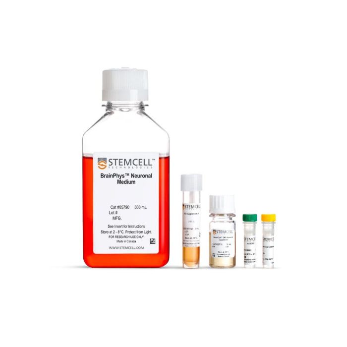
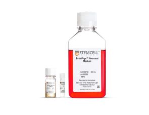
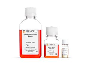
试剂盒包含 BrainPhys™ 神经元培养基、SM1 神经元补充剂、N2 补充剂-A、BDNF 和 GDNF,用于培养 ES/iPS 细胞衍生的神经元

产品优势

  • 更加贴近大脑细胞外微环境
  • 提高神经元功能,并促进突触活性神经元比例的提升
  • 无需更换培养基、避免细胞应激的情况下,直接开展功能性检测
  • 支持ES/iPS细胞及中枢神经系统(CNS)的神经元的长期培养
  • 严格的原材料筛选与质量控制,有效降低批次间差异,确保实验稳定性

产品组分包括

  • BrainPhys™ 神经元培养基,500 mL(产品号 #05790)
  • NeuroCult™ SM1 神经元添加物,10 mL(产品号 #05711)
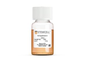
  • N2 Supplement-A,5 mL(产品号 #07152)
  • 人重组 BDNF,10 µg(产品号 #78005)
  • 人重组GDNF,10 µg(产品号 #78058)

What Our Scientist Says

I want to help neuroscientists like you create more physiological culture conditions, for more active and healthy neuronal cultures.

Carmen MakScientist
Carmen Mak, Scientist

总览

使用优化配方的完整培养基,可用于培养、分化并成熟由人胚胎干(ES)细胞或诱导多能干(iPS)细胞分化的神经元,其设计目的是促进而非抑制神经元活性。

为提升使用便利性,BrainPhys™ hPSC神经元试剂盒提供了无血清BrainPhys™神经元培养基(基础培养基)、添加物和生长因子,帮助您从人ES/iPS细胞来源的神经祖细胞中生成并成熟不同类型的神经元。

BrainPhys™ 神经元培养基基于 Bardy与Gage(Bardy et al. PNAS, 2015)的配方,模拟中枢神经系统(CNS)的细胞外环境,从而诱导更高比例的突触活性神经元。其中,基于Brewer B27配方(Brewer et al. J Neurosci Res., 1993)的NeuroCult™ SM1神经元添加物可维持细胞健康,并在短期与长期无血清培养条件下促进神经突起生长与分支;N2 Supplement-A则支持ES/iPS细胞向多种神经元亚型的分化。此外,试剂盒中还包含BDNF和GDNF生长因子,用于支持谱系特异性的分化。

为了避免因培养基更换而对细胞造成压力,您还可以在进行功能性分析(例如微电极阵列记录或实时荧光成像)时使用 BrainPhys™ 培养基。

查看我们的其他资源,了解更多关于BrainPhys™ 系统的信息。

分类
基础培养基,专用培养基
 
细胞类型
神经细胞,PSC衍生,神经元,多能干细胞
 
种属

 
应用
细胞培养,分化,培养
 
品牌
BrainPhys
 
研究领域
疾病建模,药物发现和毒理检测,神经科学,干细胞生物学
 
制剂类别
无血清
 

实验数据

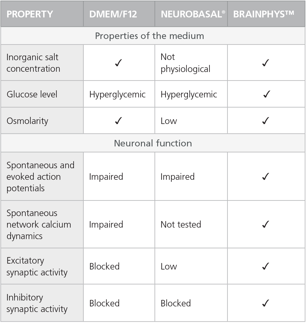
Table 1. Properties of Culture Media (C Bardy et al. Proc Natl Acad Sci USA, 2015)

Check-mark denotes physiological conditions

Check-mark denotes physiological conditions and supported activities according to C Bardy et al. Proc Natl Acad Sci USA, 2015.

Rodent Neurons Matured in BrainPhys™ Neuronal Medium

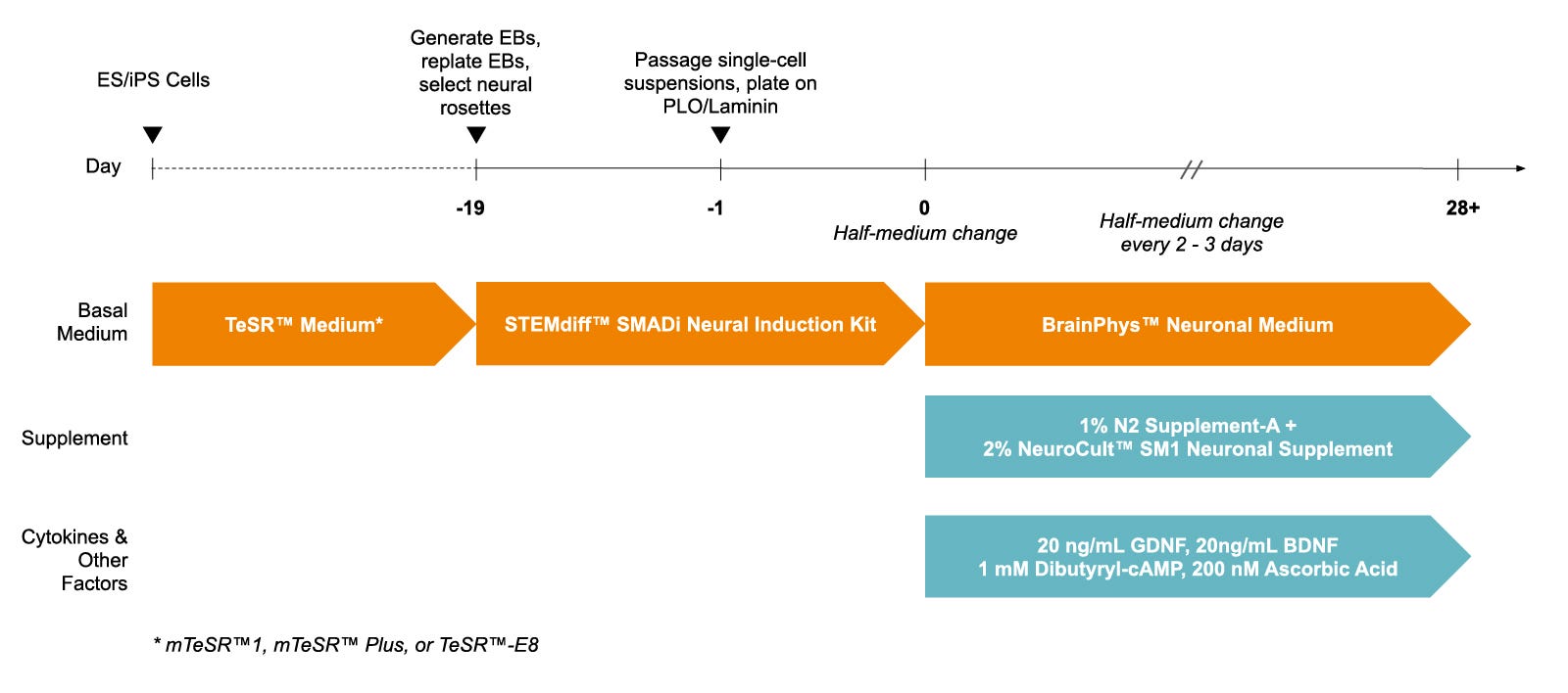
Figure 1. Protocol for Culturing hPSCs with the SM1 Culture System

hPSCs were maintained in mTeSR™1 medium and then differentiated using the STEMdiff™ SMADi Neural Induction Kit. Following plating on PLO/laminin, half-medium changes were performed to transition to BrainPhys™ Neuronal Medium for maturation and long-term culture.

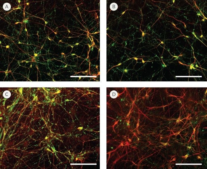
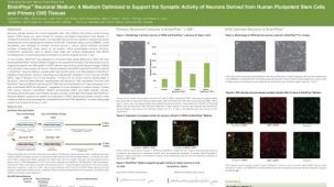
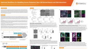
hPSC-Derived Neurons Generated in BrainPhys™ Neuronal Medium Express Markers of Neuronal Maturity After 14 and 44 Days of Differentiation

Figure 2. hPSC-Derived Neurons Generated in BrainPhys™ Neuronal Medium Express Markers of Neuronal Maturity After 14 and 44 Days of Differentiation

NPCs were generated from H9 cells using STEMdiff™ Neural Induction Medium in an embryoid body-based protocol. Next, NPCs were cultured in (A,C) BrainPhys™ Neuronal Medium, supplemented with 2% NeuroCult™ SM1 Supplement, 1% N2 Supplement-A, 20 ng/mL GDNF, 20 ng/mL BDNF, 1 mM db-cAMP and 200 nM ascorbic acid to initiate neuronal differentiation, or (B,D) DMEM/F12 under the same supplementation conditions. After 14 and 44 days of differentiation and maturation, neurons express the synaptic marker Synapsin 1 (green) and the mature neuronal marker MAP2 (red). In this example, neurons matured in BrainPhys™ Neuronal Medium show increased Synapsin 1 staining. Scale bar= 100 µm

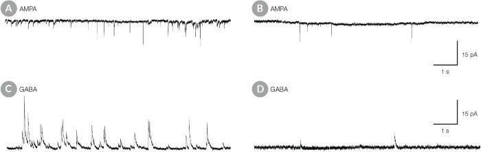
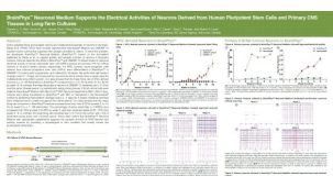
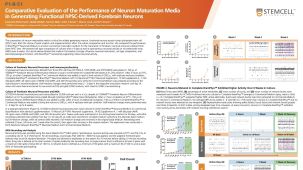
hPSC-Derived Neurons Matured in BrainPhys™ Neuronal Medium Show Improved Excitatory and Inhibitory Synaptic Activity

Figure 3. hPSC-Derived Neurons Matured in BrainPhys™ Neuronal Medium Show Improved Excitatory and Inhibitory Synaptic Activity

NPCs were generated from H9 cells using STEMdiff™ Neural Induction Medium in an embryoid body-based protocol. Next, NPCs were cultured for 44 DIV in (A,C) BrainPhys™ Neuronal Medium, supplemented with 2% NeuroCult™ SM1 Supplement, 1% N2 Supplement-A, 20 ng/mL GDNF, 20 ng/mL BDNF, 1 mM db-cAMP and 200 nM ascorbic acid to initiate neuronal differentiation, or (B,D) in DMEM/F12 under the same supplementation conditions. (A,C) Neurons matured in BrainPhys™ Neuronal Medium showed spontaneous excitatory (AMPA-mediated; A) and inhibitory (GABA-mediated; C) synaptic events. The frequency and amplitude of spontaneous synaptic events is consistently greater in neuronal cultures matured in BrainPhys™ Neuronal Medium, compared to neurons plated and matured in DMEM/F12 (B,D). Traces are representative.

产品说明书及文档

请在《产品说明书》中查找相关支持信息和使用说明,或浏览下方更多实验方案。

Document Type
Product Name
Catalog #
Lot #
Language
Catalog #
05795
Lot #
All
Language
English
Document Type
Safety Data Sheet 1
Catalog #
05795
Lot #
All
Language
English
Document Type
Safety Data Sheet 2
Catalog #
05795
Lot #
All
Language
English
Document Type
Safety Data Sheet 3
Catalog #
05795
Lot #
All
Language
English

应用领域

本产品专为以下研究领域设计,适用于工作流程中的高亮阶段。探索这些工作流程,了解更多我们为各研究领域提供的其他配套产品。

相关材料与文献

技术资料 (27)

文献 (52)

Development and Dynamic Regulation of Mitochondrial Network in Human Midbrain Dopaminergic Neurons Differentiated from iPSCs. E. Gabriel et al. Stem cell reports 2016 JAN

Abstract

Mitochondria are critical to neurogenesis,but the mechanisms of mitochondria in neurogenesis have not been well explored. We fully characterized mitochondrial alterations and function in relation to the development of human induced pluripotent stem cell (hiPSC)-derived dopaminergic (DA) neurons. Following directed differentiation of hiPSCs to DA neurons,mitochondria in these neurons exhibit pronounced changes during differentiation,including mature neurophysiology characterization and functional synaptic network formation. Inhibition of mitochondrial respiratory chains via application of complex IV inhibitor KCN (potassium cyanide) or complex I inhibitor rotenone restricted neurogenesis of DA neurons. These results demonstrated the direct importance of mitochondrial development and bioenergetics in DA neuronal differentiation. Our study also provides a neurophysiologic model of mitochondrial involvement in neurogenesis,which will enhance our understanding of the role of mitochondrial dysfunctions in neurodegenerative diseases.
A viral strategy for targeting and manipulating interneurons across vertebrate species. Dimidschstein J et al. Nature neuroscience 2016 DEC

Abstract

A fundamental impediment to understanding the brain is the availability of inexpensive and robust methods for targeting and manipulating specific neuronal populations. The need to overcome this barrier is pressing because there are considerable anatomical,physiological,cognitive and behavioral differences between mice and higher mammalian species in which it is difficult to specifically target and manipulate genetically defined functional cell types. In particular,it is unclear the degree to which insights from mouse models can shed light on the neural mechanisms that mediate cognitive functions in higher species,including humans. Here we describe a novel recombinant adeno-associated virus that restricts gene expression to GABAergic interneurons within the telencephalon. We demonstrate that the viral expression is specific and robust,allowing for morphological visualization,activity monitoring and functional manipulation of interneurons in both mice and non-genetically tractable species,thus opening the possibility to study GABAergic function in virtually any vertebrate species.
Recent Zika Virus Isolates Induce Premature Differentiation of Neural Progenitors in Human Brain Organoids. E. Gabriel et al. Cell stem cell 2017 JAN

Abstract

The recent Zika virus (ZIKV) epidemic is associated with microcephaly in newborns. Although the connection between ZIKV and neurodevelopmental defects is widely recognized,the underlying mechanisms are poorly understood. Here we show that two recently isolated strains of ZIKV,an American strain from an infected fetal brain (FB-GWUH-2016) and a closely-related Asian strain (H/PF/2013),productively infect human iPSC-derived brain organoids. Both of these strains readily target to and replicate in proliferating ventricular zone (VZ) apical progenitors. The main phenotypic effect was premature differentiation of neural progenitors associated with centrosome perturbation,even during early stages of infection,leading to progenitor depletion,disruption of the VZ,impaired neurogenesis,and cortical thinning. The infection pattern and cellular outcome differ from those seen with the extensively passaged ZIKV strain MR766. The structural changes we see after infection with these more recently isolated viral strains closely resemble those seen in ZIKV-associated microcephaly.

更多信息

更多信息
物种
配方 无血清
法律声明:

BrainPhys is a registered trademark of the Salk Institute for Biological Studies, used under exclusive license. 质量保证:

产品仅供研究使用,不用于针对人或动物的诊断或治疗。 欲获悉更多关于STEMCELL的质控信息,请访问 STEMCELL.CN/COMPLIANCE.
Copyright © 2026 by STEMCELL Technologies. All rights reserved.

在线联系