产品号 #08571_C
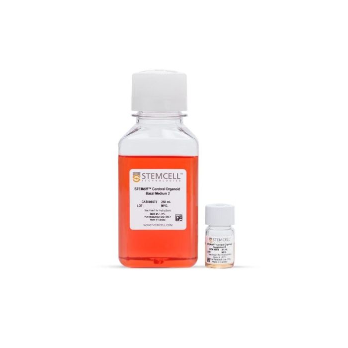
用于人脑类器官成熟的培养基试剂盒
若您需要咨询产品或有任何技术问题,请通过官方电话 400 885 9050 或邮箱 info.cn@stemcell.com 与我们联系。
用于人脑类器官成熟的培养基试剂盒
用于人脑类器官成熟的培养基试剂盒
Human brain development is complex, so to observe it in a dish is a huge scientific breakthrough. We've tried to simplify that process and make it more accessible to you, regardless of how much stem cell experience you have.

用STEMdiff™ 脑类器官成熟试剂盒培养多能干细胞(PSC)来源的神经类器官,可延长培养周期(>40天)。
为方便使用,该成熟试剂盒与STEMdiff™ 脑类器官试剂盒完全兼容,后者可在明确的无血清条件下生成神经类器官。
如需了解STEMdiff™ 脑类器官成熟试剂盒培养类器官的实验方案,请查阅产品说明书(PIS)及教育资料。
有关使用 STEMdiff™ 脑类器官成熟试剂盒进行培养类器官的更多方案信息,请参阅产品说明书(PIS)及技术资料。
分类
专用培养基
细胞类型
神经细胞,PSC衍生,神经干/祖细胞,多能干细胞
种属
人
应用
细胞培养,鉴定,分化,功能学筛选,免疫荧光,类器官培养,表型鉴定,球状体培养
品牌
STEMdiff
研究领域
疾病建模,神经科学,干细胞生物学
制剂类别
无血清

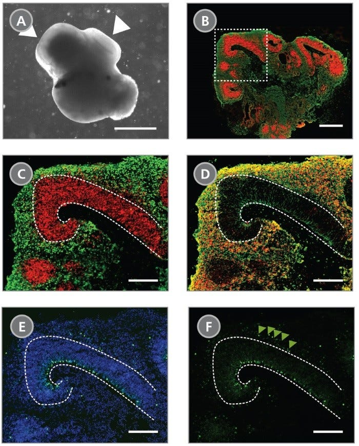
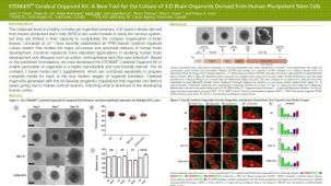
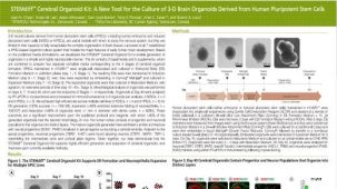
Figure 1. Cerebral Organoids Contain Multiple Layered Regions That Recapitulate the Cortical Lamination Process Observed During In Vivo Human Brain Development
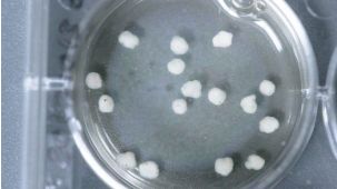
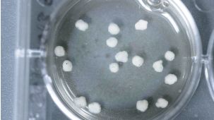
(A) A representative phase-contrast image of a whole cerebral organoid at Day 40 generated using the STEMdiff™ Cerebral Organoid Kit. Cerebral organoids at this stage are made up of phase-dark structures that may be surrounded by regions of thinner, more translucent structures that display layering (arrowheads). (B) Immunohistological analysis on cryosections of cerebral organoids reveals cortical regions within the organoid labeled by the apical progenitor marker PAX6 (red) and neuronal marker β-tubulin III+ (TUJ-1) (green). (C-F) Inset of boxed region from (B). (C) PAX6+ apical progenitors (red, enclosed by dotted line) are localized to a ventricular zone-like region. β-tubulin III+ neurons (green) are adjacent to the ventricular zone. (D) CTIP2, a marker of the developing cortical plate, co-localizes with β-tubulin III+ neurons in a cortical plate-like region. Organization of the layers recapitulates early corticogenesis observed during human brain development. (E) Proliferating progenitor cells labeled by Ki-67 (green) localize along the ventricle, nuclei are counterstained with DAPI (blue). (F) An additional population of Ki-67+ cells is found in an outer subventricular zone-like region (arrowheads). Scale bar = (A) 1 mm, (B) 1 mm and (C-F) 200 µm.

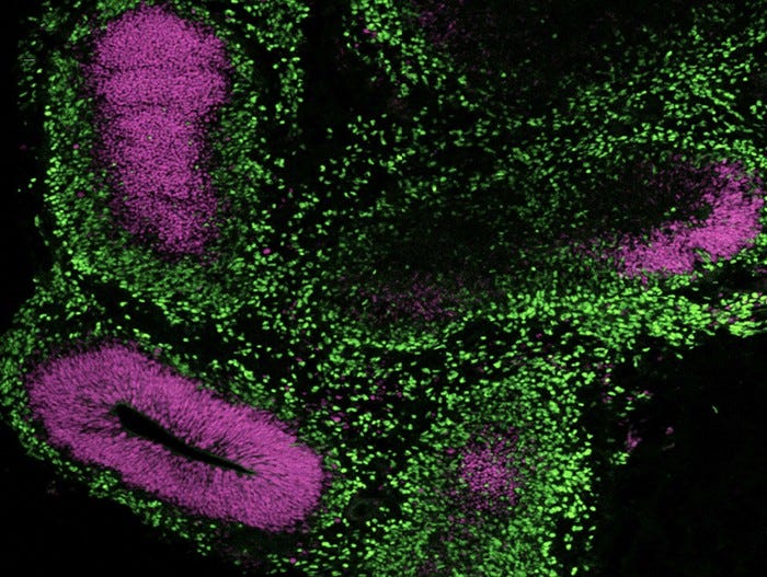
Figure 2. Cerebral Organoids Can Be Generated from hPSCs Maintained in mTeSR™ Plus
Human ES (H9) cells were cultured with mTeSR™ Plus and directed to cerebral organoids using the STEMdiff™ Cerebral Organoid Kit. Image shows apical progenitor marker SOX2 (magenta) and neuronal marker TBR1 (green).

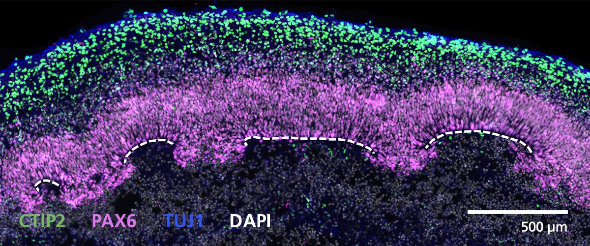
Figure 3. Cryosectioned Cerebral Organoids Show Stratification of Cortical Plate Neurons and Progenitor Zones
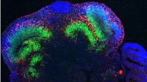
Cerebral organoids were generated using the STEMdiff™ Cerebral Organoid Kit. A 16-μm-thick section of a Day 40 cerebral organoid was stained for CTIP2 (green), PAX6 (magenta), βIII-tubulin/TUJ1 (blue), and DAPI (gray). Cortical regions are defined by progenitor cells (PAX6+) that are radially organized around a pseudo-ventricle (dashed line). These progenitors give rise to cortical plate neurons indicated by CTIP2 and TUJ1 expression. For a detailed cryogenic tissue processing and immunofluorescence protocol, please see the Methods Library.

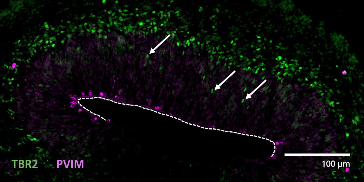
Figure 4. Zones of Active Proliferation in Cerebral Organoids Are Preserved Following the Protocol for Tissue Processing
Organoid tissue was processed for immunofluorescence and stained for TBR2 (intermediate precursors, green) and phosphorylated vimentin (PVIM, dividing cells, magenta). Cells actively divide at the apical border of cortical regions along the border of the pseudo-ventricle (dashed line). A population of these dividing cells will express TBR2 and then migrate (arrows) from the progenitor zone to form a layer of intermediate progenitors. For a detailed cryogenic tissue processing and immunofluorescence protocol, please see the Methods Library.

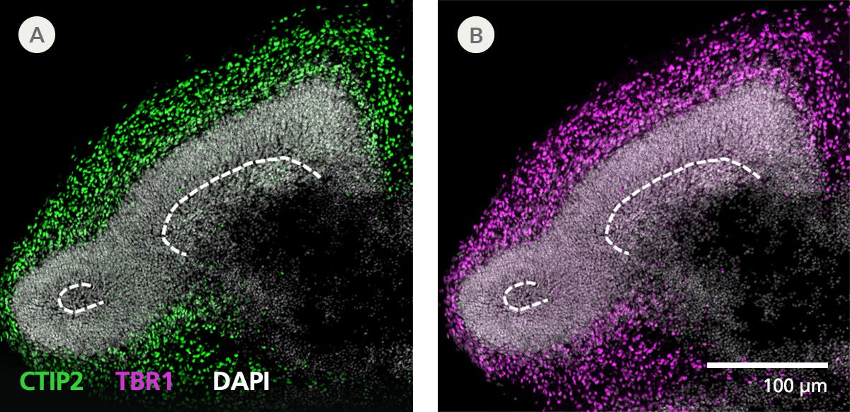
Figure 5. Immunofluorescence from Cryosectioned Cerebral Organoids Indicates Preserved Organization of Cortical Neurons
Organoid tissue was processed for immunofluorescence and stained for CTIP2 (green), TBR1 (layer 5/6 cortical neurons, magenta), and DAPI (white). Deep layer neuronal markers CTIP2 and TBR1 are expressed in cells around presumptive progenitor zones (dashed line) toward the outside or apical surface of organoids. For a detailed cryogenic tissue processing and immunofluorescence protocol, please see the Methods Library.

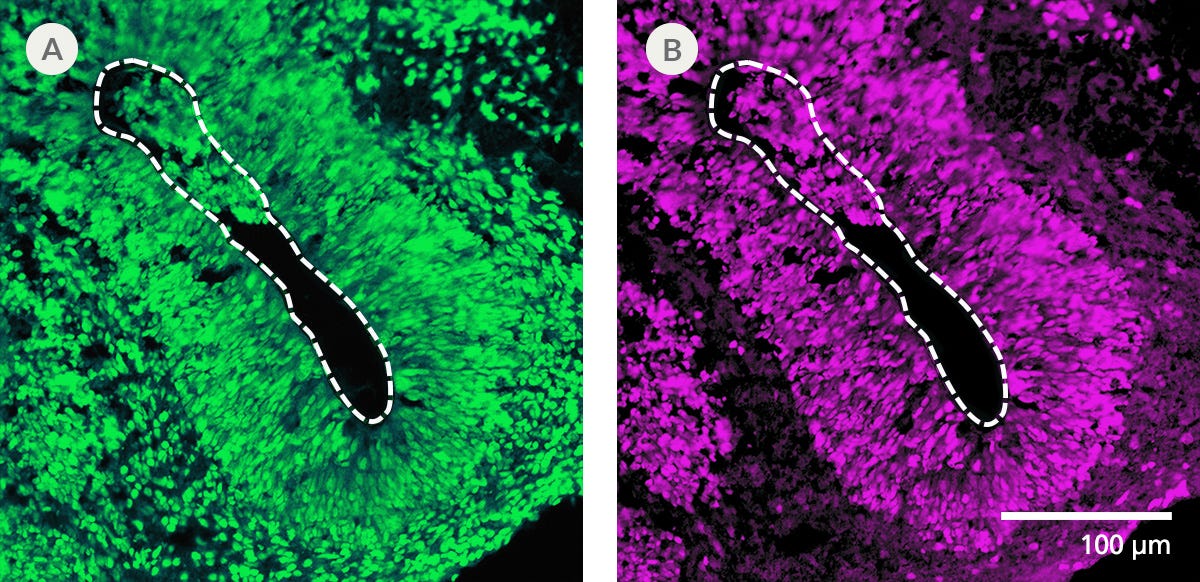
Figure 6. Cryogenic Tissue Processing and Immunofluorescence Captures Arrangement of Neural Progenitors Around Pseudo-Ventricles in Cerebral Organoids
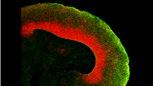
Organoid tissue was processed for immunofluorescence and stained for (A) FOXG1 (forebrain cells, green) or (B) SOX2 (neural progenitors, magenta). Organoids derived from STEMdiff™ Cerebral Organoid Kit generate forebrain-type tissue as indicated by FOXG1 expression. Neural progenitors expressing SOX2 are radially arranged around a pseudo-ventricle area (dashed line). For a detailed cryogenic tissue processing and immunofluorescence protocol, please see the Methods Library.

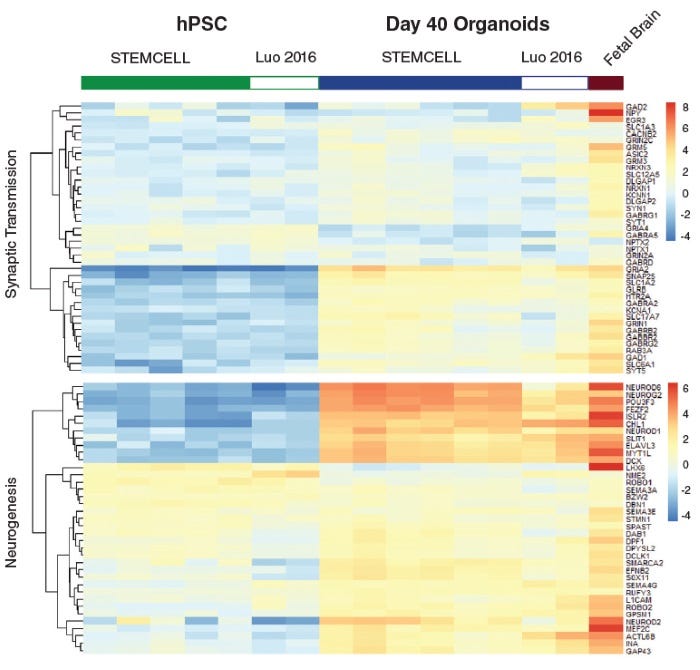
Figure 7. Neural Organoids Generated with STEMdiff™ Cerebral Organoid Kit Express Expected Key Markers
Heatmap of expression levels for genes associated with synaptic transmission function and neurogenesis in Day 40 organoids. These data show that gene expression of cerebral organoids generated from the STEMdiff™ Cerebral Organoid Kit are similar to published results (C Luo et al. Cell Rep, 2016).

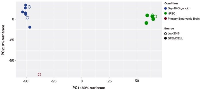
Figure 8. Cerebral Organoids Generated with the STEMdiff™ Cerebral Organoid Kit Are Transcriptionally Similar to Those from Published Protocols
Principal component analysis of hPSC and cerebral organoid transcriptomes. Cerebral organoids generated using the STEMdiff™ Cerebral Organoid Kit (filled blue circles) cluster together, and cluster with previously published (C Luo et al. Cell Rep, 2016) cerebral organoids (open blue circles). The first principal component accounts for the majority of variance seen (PC1; 80%) and distinguishes the cerebral organoid samples from the hPSCs (green circles). The second principal component accounts for only 9% of the variation, and highlights the modest expression differences between cultured organoids and primary embryonic fetal brain samples (19 post-conceptional weeks, brown circles).

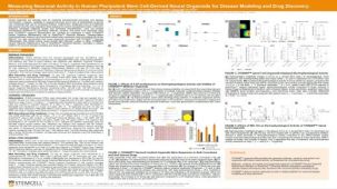
Figure 9. STEMdiff™ Cerebral Organoids Exhibit Synchronous Calcium Activity
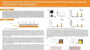
Cerebral organoids were generated using the STEMdiff™ Cerebral Organoid Kit and matured with the STEMdiff™ Cerebral Organoid Maturation Kit. On Day 91, organoids were imaged using the ImageXpress® Confocal HT.ai High-Content Imaging System (Molecular Devices) with MetaXpress® High-Content Image Acquisition and Analysis Software. (A) Time-lapse images of a representative region within a cerebral organoid. Arrows indicate areas of elevated calcium signal intensity. (B) Corresponding heatmap representation of calcium intensity from the regions shown in (A) over time. Signal propagation from the initial peak to surrounding areas suggests the presence of a functional neuronal network.
请在《产品说明书》中查找相关支持信息和使用说明,或浏览下方更多实验方案。
本产品专为以下研究领域设计,适用于工作流程中的高亮阶段。探索这些工作流程,了解更多我们为各研究领域提供的其他配套产品。
Thank you for your interest in IntestiCult™ Organoid Growth Medium (Human). Please provide us with your contact information and your local representative will contact you with a customized quote. Where appropriate, they can also assist you with a(n):
Estimated delivery time for your area
Product sample or exclusive offer
In-lab demonstration
| 物种 | 人 |
|---|---|
| 配方 | 无血清 |
提升神经元功能的无血清基础培养基
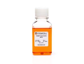
用于人 ES 和 iPS 细胞神经诱导的成分明确的无血清培养基
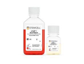
<p>cGMP标准、无饲养层的hESC和iPSC维持培养基</p>
Legal Statement:
This product was developed under a license to intellectual property owned by the Institute of Molecular Biotechnology (IMBA) of the Austrian Academy of Sciences. This product is sold for research use only (whether the buyer is an academic or for-profit entity) under a non-transferable, limited-use license. Purchase of this product does not include the right to sell, use or otherwise transfer this product for commercial purposes (i.e., any activity undertaken for consideration, such as use of this product for manufacturing, or resale of this product or any materials made using this product, or use of this product or any materials made using this product to provide services or, in collaboration with, a for-profit entity, for purposes other than research applications (i.e., drug development activities). Purchasers wishing to use the product for commercial purposes should contact IMBA at technologvtransfer@imba.oeaw.ac.at.
Quality Statement:
PRODUCTS ARE FOR RESEARCH USE ONLY AND NOT INTENDED FOR HUMAN OR ANIMAL DIAGNOSTIC OR THERAPEUTIC USES UNLESS OTHERWISE STATED. FOR ADDITIONAL INFORMATION ON QUALITY AT STEMCELL, REFER TO WWW.STEMCELL.CN/COMPLIANCE.
Safety Statement:
CA WARNING: This product can expose you to Progesterone which is known to the State of California to cause cancer. For more information go to www.P65Warnings.ca.gov
扫描二维码或搜索微信号STEMCELLTech,即可关注我们的微信平台,第一时间接收丰富的技术资源和最新的活动信息。
如您有任何问题,欢迎发消息给STEMCELLTech微信公众平台,或与我们通过电话/邮件联系:400 885 9050 INFO.CN@STEMCELL.COM。
在线联系