Dalley AJ et al. (JAN 2013)
Journal of oral pathology & medicine : official publication of the International Association of Oral Pathologists and the American Academy of Oral Pathology 42 1 37--46
Organotypic culture of normal, dysplastic and squamous cell carcinoma-derived oral cell lines reveals loss of spatial regulation of CD44 and p75 NTR in malignancy.
Oral squamous cell carcinomas (OSCC) often arise from dysplastic lesions. The role of cancer stem cells in tumour initiation is widely accepted,yet the potential existence of pre-cancerous stem cells in dysplastic tissue has received little attention. Cell lines from oral diseases ranging in severity from dysplasia to malignancy provide opportunity to investigate the involvement of stem cells in malignant progression from dysplasia. Stem cells are functionally defined by their ability to generate hierarchical tissue structures in consortium with spatial regulation. Organotypic cultures readily display tissue hierarchy in vitro; hence,in this study,we compared hierarchical expression of stem cell-associated markers in dermis-based organotypic cultures of oral epithelial cells from normal tissue (OKF6-TERT2),mild dysplasia (DOK),severe dysplasia (POE-9n) and OSCC (PE/CA P J15). Expression of CD44,p75(NTR),CD24 and ALDH was studied in monolayers by flow cytometry and in organotypic cultures by immunohistochemistry. Spatial regulation of CD44 and p75(NTR) was evident for organotypic cultures of normal (OKF6-TERT2) and dysplasia (DOK and POE-9n) but was lacking for OSCC (PE/CA PJ15)-derived cells. Spatial regulation of CD24 was not evident. All monolayer cultures exhibited CD44,p75(NTR),CD24 antigens and ALDH activity (ALDEFLUOR(®) assay),with a trend towards loss of population heterogeneity that mirrored disease severity. In monolayer,increased FOXA1 and decreased FOXA2 expression correlated with disease severity,but OCT3/4,Sox2 and NANOG did not. We conclude that dermis-based organotypic cultures give opportunity to investigate the mechanisms that underlie loss of spatial regulation of stem cell markers seen with OSCC-derived cells.
View Publication
产品号#:
01700
01705
01702
产品名:
ALDEFLUOR™ 试剂盒
ALDEFLUOR™ DEAB试剂, 1.5 mM, 1 mL
ALDEFLUOR™检测缓冲液
Okkelman IA et al. ( 2016)
PloS one 11 12 e0167385
Use of Fluorescence Lifetime Imaging Microscopy (FLIM) as a Timer of Cell Cycle S Phase.
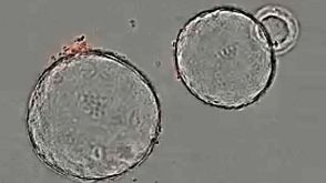
Incorporation of thymidine analogues in replicating DNA,coupled with antibody and fluorophore staining,allows analysis of cell proliferation,but is currently limited to monolayer cultures,fixed cells and end-point assays. We describe a simple microscopy imaging method for live real-time analysis of cell proliferation,S phase progression over several division cycles,effects of anti-proliferative drugs and other applications. It is based on the prominent (˜ 1.7-fold) quenching of fluorescence lifetime of a common cell-permeable nuclear stain,Hoechst 33342 upon the incorporation of 5-bromo-2'-deoxyuridine (BrdU) in genomic DNA and detection by fluorescence lifetime imaging microscopy (FLIM). We show that quantitative and accurate FLIM technique allows high-content,multi-parametric dynamic analyses,far superior to the intensity-based imaging. We demonstrate its uses with monolayer cell cultures,complex 3D tissue models of tumor cell spheroids and intestinal organoids,and in physiological study with metformin treatment.
View Publication

 EasySep™小鼠TIL(CD45)正选试剂盒


 沪公网安备31010102008431号