若您需要咨询产品或有任何技术问题,请通过官方电话 400 885 9050 或邮箱 info.cn@stemcell.com 与我们联系。

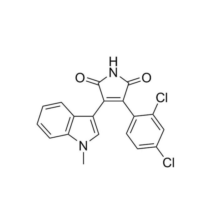
SB216763

WNT通路激活剂;抑制GSK3α和GSK3β
只有 %1
¥1,438.00

产品号 #(选择产品)

产品号 #72872_C

WNT通路激活剂;抑制GSK3α和GSK3β

总览

SB216763 是一种可透过细胞膜的 ATP 竞争性糖原合酶激酶 3α(GSK3α,IC₅₀ = 34 nM)及同工酶 GSK3β 的抑制剂(Coghlan et al.)。GSK3是一种丝氨酸/苏氨酸蛋白激酶,会被多种细胞外刺激的抑制,如胰岛素、生长因子、细胞命运决定因子和细胞粘附。

维持和自我更新
·在无白血病抑制因子(LIF)的情况下,将小鼠胚胎干细胞(ES)与小鼠胚胎成纤维细胞(MEFs)共培养,维持在未分化的多能状态长达两个月(Kirby et al.)。
·促进原代小鼠视网膜干细胞的增殖(Inoue et al.)。
·增加小鼠大脑中的神经祖细胞增殖(Mao et al.)。
·在成年小鼠的体内和体外模型中,促进神经干细胞(NSCs)的对称分裂(Piccin and Morshead)。
·抑制人间充质干细胞(MSCs)向脂肪细胞的分化(Shen et al.)。
·促进主动脉-性腺-中肾(AGM)外植体培养中造血干细胞(HSC)的生成(Ruiz-Herguido et al.)。

分化
·增强胰岛素诱导的小鼠肌母细胞静息储备细胞的分化(Rochat et al.)。
·刺激大鼠神经球的NSC的分化(Maurer et al.)。
·诱导人神经祖细胞(NPC)向神经元分化(Lange et al.)。
·促进小鼠造血祖细胞(HPCs)向树突状细胞分化(Zhou et al.)。

癌症研究
·诱导人胶质母细胞瘤细胞分化并减少癌症干细胞群(Korur et al.)。

细胞类型
癌细胞及细胞系,树突状细胞(DCs),造血干/祖细胞,间充质干/祖细胞,肌源干/祖细胞,神经干/祖细胞,多能干细胞
 
种属
人,小鼠,非人灵长类,其他物种,大鼠
 
应用
分化,扩增,培养
 
研究领域
癌症,神经科学,干细胞生物学
 
CAS 编号
280744-09-4
 
化学式
C₁₉H₁₂Cl₂N₂O₂
 
纯度
≥98%
 
通路
WNT
 
靶点
GSK
 

产品说明书及文档

请在《产品说明书》中查找相关支持信息和使用说明,或浏览下方更多实验方案。

Document Type
Product Name
Catalog #
Lot #
Language
Product Name
SB216763
Catalog #
72872
Lot #
All
Language
English
Document Type
Safety Data Sheet
Product Name
SB216763
Catalog #
72872
Lot #
All
Language
English

相关材料与文献

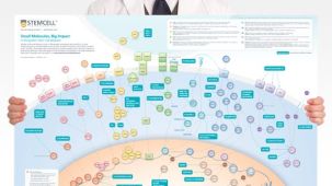
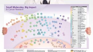
技术资料 (3)

文献 (12)

Selective small molecule inhibitors of glycogen synthase kinase-3 modulate glycogen metabolism and gene transcription. Coghlan MP et al. Chemistry & biology 2000 OCT

Abstract

BACKGROUND Glycogen synthase kinase-3 (GSK-3) is a serine/threonine protein kinase,the activity of which is inhibited by a variety of extracellular stimuli including insulin,growth factors,cell specification factors and cell adhesion. Consequently,inhibition of GSK-3 activity has been proposed to play a role in the regulation of numerous signalling pathways that elicit pleiotropic cellular responses. This report describes the identification and characterisation of potent and selective small molecule inhibitors of GSK-3. RESULTS SB-216763 and SB-415286 are structurally distinct maleimides that inhibit GSK-3alpha in vitro,with K(i)s of 9 nM and 31 nM respectively,in an ATP competitive manner. These compounds inhibited GSK-3beta with similar potency. However,neither compound significantly inhibited any member of a panel of 24 other protein kinases. Furthermore,treatment of cells with either compound stimulated responses characteristic of extracellular stimuli that are known to inhibit GSK-3 activity. Thus,SB-216763 and SB-415286 stimulated glycogen synthesis in human liver cells and induced expression of a beta-catenin-LEF/TCF regulated reporter gene in HEK293 cells. In both cases,compound treatment was demonstrated to inhibit cellular GSK-3 activity as assessed by activation of glycogen synthase,which is a direct target of this kinase. CONCLUSIONS SB-216763 and SB-415286 are novel,potent and selective cell permeable inhibitors of GSK-3. Therefore,these compounds represent valuable pharmacological tools with which the role of GSK-3 in cellular signalling can be further elucidated. Furthermore,development of similar compounds may be of use therapeutically in disease states associated with elevated GSK-3 activity such as non-insulin dependent diabetes mellitus and neurodegenerative disease.
Insulin and wnt1 pathways cooperate to induce reserve cell activation in differentiation and myotube hypertrophy. Rochat A et al. Molecular biology of the cell 2004 OCT

Abstract

During ex vivo myoblast differentiation,a pool of quiescent mononucleated myoblasts,reserve cells,arise alongside myotubes. Insulin/insulin-like growth factor (IGF) and PKB/Akt-dependent phosphorylation activates skeletal muscle differentiation and hypertrophy. We have investigated the role of glycogen synthase kinase 3 (GSK-3) inhibition by protein kinase B (PKB)/Akt and Wnt/beta-catenin pathways in reserve cell activation during myoblast differentiation and myotube hypertrophy. Inhibition of GSK-3 by LiCl or SB216763,restored insulin-dependent differentiation of C2ind myoblasts in low serum,and cooperated with insulin in serum-free medium to induce MyoD and myogenin expression in C2ind myoblasts,quiescent C2 or primary human reserve cells. We show that LiCl treatment induced nuclear accumulation of beta-catenin in C2 myoblasts,thus mimicking activation of canonical Wnt signaling. Similarly to the effect of GSK-3 inhibitors with insulin,coculturing C2 reserve cells with Wnt1-expressing fibroblasts enhanced insulin-stimulated induction of MyoD and myogenin in reserve cells. A similar cooperative effect of LiCl or Wnt1 with insulin was observed during late ex vivo differentiation and promoted increased size and fusion of myotubes. We show that this synergistic effect on myotube hypertrophy involved an increased fusion of reserve cells into preexisting myotubes. These data reveal insulin and Wnt/beta-catenin pathways cooperate in muscle cell differentiation through activation and recruitment of satellite cell-like reserve myoblasts.
Activation of canonical Wnt pathway promotes proliferation of retinal stem cells derived from adult mouse ciliary margin. Inoue T et al. Stem cells (Dayton,Ohio) 2006 JAN

Abstract

Adult retinal stem cells represent a possible cell source for the treatment of retinal degeneration. However,only a small number of stem cells reside in the ciliary margin. The present study aimed to promote the proliferation of adult retinal stem cells via the Wnt signaling pathway. Ciliary margin cells from 8-week-old mice were dissociated and cultured to allow sphere colony formation. Wnt3a,a glycogen synthase kinase (GSK) 3 inhibitor,fibroblast growth factor (FGF) 2,and a FGF receptor inhibitor were then applied in the culture media. The primary spheres were dissociated to prepare either monolayer or secondary sphere cultures. Wnt3a increased the size of the primary spheres and the number of Ki-67-positive proliferating cells in monolayer culture. The Wnt3a-treated primary sphere cells were capable of self-renewal and gave rise to fourfold the number of secondary spheres compared with nontreated sphere cells. These cells also retained their multilineage potential to express several retinal markers under differentiating culture conditions. The Wnt3a-treated cells showed nuclear accumulation of beta-catenin,and a GSK3 inhibitor,SB216763,mimicked the mitogenic activity of Wnt3a. The proliferative effect of SB216763 was attenuated by an FGF receptor inhibitor but was enhanced by FGF2,with Ki-67-positive cells reaching over 70% of the total cells. Wnt3a and SB216763 promoted the proliferation of retinal stem cells,and this was partly dependent on FGF2 signaling. A combination of Wnt and FGF signaling may provide a therapeutic strategy for in vitro expansion or in vivo activation of adult retinal stem cells.

更多信息

更多信息
物种 人, 其它物种, 大鼠, 小鼠, 非人灵长类
Cas Number 280744-09-4
Chemical Formula C₁₉H₁₂Cl₂N₂O₂
纯度 ≥ 98%
Target GSK
Pathway WNT
质量保证:

产品仅供研究使用,不用于针对人或动物的诊断或治疗。 欲获悉更多关于STEMCELL的质控信息,请访问 STEMCELL.CN/COMPLIANCE.
Copyright © 2026 by STEMCELL Technologies. All rights reserved.

在线联系