
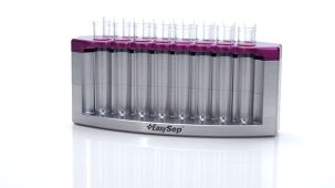
 EasySep™小鼠TIL(CD45)正选试剂盒


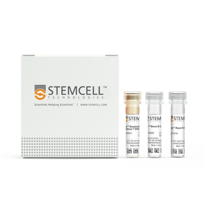
产品号 #19854_C
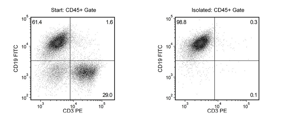
通过免疫磁珠负选分离出无磁珠标记的小鼠B细胞
若您需要咨询产品或有任何技术问题,请通过官方电话 400 885 9050 或邮箱 info.cn@stemcell.com 与我们联系。
通过免疫磁珠负选分离出无磁珠标记的小鼠B细胞
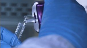
使用 EasySep™ 小鼠 B 细胞分选试剂盒,可通过免疫磁珠负选法从脾细胞或其他组织样本的单细胞悬液中轻松高效地分离出高纯度的小鼠 B 细胞。EasySep™ 技术在已发表研究中被广泛应用超过 20 年,结合了单克隆抗体的特异性与无柱磁珠分选系统的简便性。
在此 EasySep™ 负选步骤中,不需要的细胞通过抗体复合物和磁珠进行标记。表达以下标志物的细胞将被去除:CD11b、CD4、CD8a、Ly6G/C、Ter119、CD43、CD49b 和 CD90.2。经 EasySep™ 磁极分离后,带有磁珠标记的非目的细胞被去除,未标记的目标 B 细胞通过倾倒或移液方式转移至新试管中。磁珠细胞分选最快可在 15 分钟内完成,目标 B 细胞可直接用于流式细胞术、培养及基于细胞的实验等下游应用。
如需分选表达 CD11b 或 CD43 的 B 细胞,建议使用 EasySep™ 小鼠 pan-B 细胞分选试剂盒(产品号 #19844)。
了解更多关于 EasySep™ 免疫磁珠分选技术的工作原理,或了解如何使用 RoboSep™ 实现免疫磁珠细胞分选的全自动化。探索更多优化实验流程的产品,包括培养基、添加物、抗体等。
磁极兼容性
• EasySep™磁极(产品号 #18000)
• “The Big Easy” EasySep™磁极(产品号 #18001)
• EasyPlate™ EasySep™磁极(产品号 #18102)
• EasyEights™ EasySep™磁极(产品号 #18103)
• RoboSep™-S(产品号 #21000)
分类
细胞分选试剂盒
细胞类型
B 细胞
种属
小鼠
样本来源
其他组织,脾脏
分选方法
负选
应用
细胞分选
品牌
EasySep,RoboSep
研究领域
免疫
请在《产品说明书》中查找相关支持信息和使用说明,或浏览下方更多实验方案。
本产品专为以下研究领域设计,适用于工作流程中的高亮阶段。探索这些工作流程,了解更多我们为各研究领域提供的其他配套产品。
| 物种 | 小鼠 |
|---|---|
| Magnet Compatibility | • EasySep™ Magnet (Catalog #18000) • “The Big Easy” EasySep™ Magnet (Catalog #18001) • EasyPlate™ EasySep™ Magnet (Catalog 18102) • EasyEights™ EasySep™ Magnet (Catalog #18103) • RoboSep™-S (Catalog #21000) |
| 样本来源 | 其它细胞系, 脾脏 |
| Selection Method | Negative |
哺乳动物有核细胞手动计数试剂
计数板含有2个盖玻片
免疫磁珠负选人B细胞
免疫磁珠正选小鼠CD19+细胞分选试剂盒
免疫磁珠负选未标记的小鼠Pan-B(CD19+、CD19+CD138+、CD138+)细胞

细胞分选缓冲液
抗小鼠CD19的大鼠Monoclonal IgG2a抗体
质量声明:
产品仅供研究使用,不用于针对人或动物的诊断或治疗。 欲获悉更多关于STEMCELL的质控信息,请访问 STEMCELL.CN/COMPLIANCE.
在线联系


 沪公网安备31010102008431号