
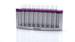
 EasySep™小鼠TIL(CD45)正选试剂盒


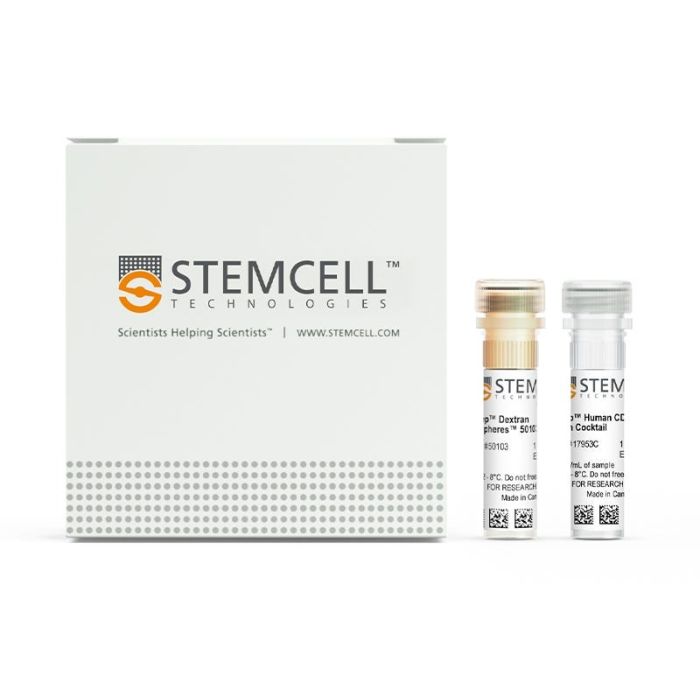
产品号 #17953_C
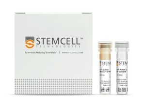
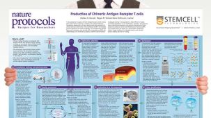
人CD8+ T细胞的免疫磁珠负选
若您需要咨询产品或有任何技术问题,请通过官方电话 400 885 9050 或邮箱 info.cn@stemcell.com 与我们联系。
人CD8+ T细胞的免疫磁珠负选
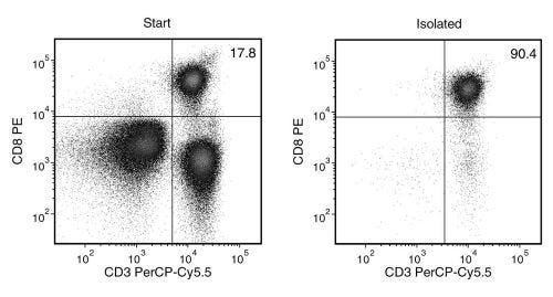
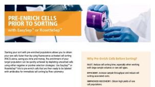
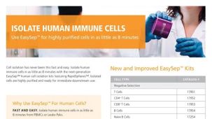
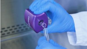
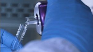
使用 EasySep™ 人 CD8⁺ T 细胞分选试剂盒,通过免疫磁性负选,可轻松高效地从新鲜或冻存的人外周血单个核细胞(PBMCs)或洗涤的白细胞单采样本中分离高纯度的人 CD8⁺ T 细胞。EasySep™ 技术结合单克隆抗体的特异性与无柱磁系统的简便性,已在发表的研究中广泛应用超过 20 年。
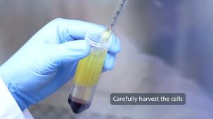
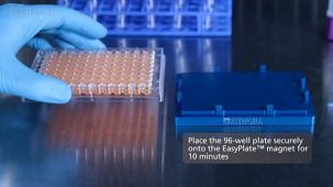
在此 EasySep™ 负选步骤中,不需要的细胞通过抗体复合物和磁珠标记。表达以下标记的非目的细胞将被去除:CD4、CD14、CD16、CD19、CD20、CD36、CD56、CD66b、CD123、GlyA 和 TCRγδ。使用 EasySep™ 磁极进行无柱分选,磁珠标记的细胞被去除,只需将未标记的CD8⁺ T 细胞倒入或移取至新的管中即可。仅需 8 分钟即可完成磁珠分选过程,分离后的CD8⁺ T细胞可直接用于下游应用,例如流式细胞术、细胞培或DNA/RNA 提取。
本产品可替代 EasySep™ 人 CD8⁺ T 细胞富集试剂盒(产品号 #19053),实现更快速的细胞分选。
如需从白细胞单采术样本中进行大规模(1×10¹⁰ 个细胞)的人 CD8⁺ T 细胞分离,请参阅大规格试剂盒(产品号 #100-0710)。
了解更多关于 EasySep™ 免疫磁性技术的工作原理,或了解如何使用 RoboSep™ 实现全自动化的免疫磁珠细胞分选。您也可以选择使用 EasySep™ 人 CD8⁺ T 细胞分选试剂盒制备的即用型、符合伦理的冻存人外周血 CD8⁺ T 细胞。探索更多优化实验流程的产品,包括培养基、补充物、抗体等。
磁极兼容性
• EasySep™磁极(产品号 #18000)
• “The Big Easy” EasySep™磁极(产品号 #18001)
• EasyPlate™ EasySep™磁极(产品号 #18102)
• Easy 50 EasySep™磁极(产品号 #18002)
• EasyEights™ EasySep™磁极(产品号 #18103)
• RoboSep™-S(产品号 #21000)
• Easy 250 EasySep™磁极(产品号 #100-0821)
分类
细胞分选试剂盒
细胞类型
T 细胞,T 细胞,CD8+
种属
人
样本来源
白细胞单采术样本、PBMC
分选方法
负选
应用
细胞分选
品牌
EasySep,RoboSep
研究领域
免疫
请在《产品说明书》中查找相关支持信息和使用说明,或浏览下方更多实验方案。
本产品专为以下研究领域设计,适用于工作流程中的高亮阶段。探索这些工作流程,了解更多我们为各研究领域提供的其他配套产品。
| 物种 | 人 |
|---|---|
| Magnet Compatibility | • EasySep™ Magnet (Catalog #18000) • “The Big Easy” EasySep™ Magnet (Catalog #18001) • Easy 50 EasySep™ Magnet (Catalog #18002) • EasyPlate™ EasySep™ Magnet (Catalog 18102) • EasyEights™ EasySep™ Magnet (Catalog #18103) • RoboSep™-S (Catalog #21000) • Eas |
| 样本来源 | PBMC, 白细胞单采术样本 |
| Selection Method | Negative |
细胞解离试剂
人CD8+ T细胞的免疫磁珠正选

冻存的人原代细胞
小鼠Monoclonal IgG1抗体,抗人、恒河猴、食蟹猴CD8a
小鼠(BALB/c)Monoclonal IgG1抗体,抗人、黑猩猩CD3
质量保证:
本产品仅供科研使用,除非另有说明,不得用于人体或动物的诊断或治疗用途。如需了解 STEMCELL 的质量体系,请访问 WWW.STEMCELL.CN/COMPLIANCE
在线联系


 沪公网安备31010102008431号