Bruserud &O et al. (APR 2004)
Haematologica 89 4 391--402
Osteoblasts increase proliferation and release of pro-angiogenic interleukin 8 by native human acute myelogenous leukemia blasts.
BACKGROUND AND OBJECTIVES: Interactions between acute myelogenous leukemia (AML) blasts and non-leukemic cells in the bone marrow seem to be important for both disease development and susceptibility to chemotherapy. Recent studies have focused on the endothelial cells,but other non-leukemic cells may also be involved. In the present study we investigated how osteoblasts affect native human AML blasts. DESIGN AND METHODS: AML cells were derived from a large group of consecutive patients. The AML blasts and osteoblastic sarcoma cell lines (Cal72,SJSA-1) were incubated together in different chambers separated by a semipermeable membrane. We investigated effects of co-culture on proliferation,apoptosis and cytokine release. RESULTS: The cross-talk between these two cell populations,achieved via release of soluble mediators,resulted in increased AML blast proliferation,including increased proliferation of clonogenic progenitors,but did not affect spontaneous in vitro apoptosis. Both interleukin (IL) 1-b and granulocyte-macrophage colony-stimulating factor were involved in this growth-enhancing cross-talk,and normal osteoblasts could also increase the AML blast proliferation. Furthermore,co-culture of AML blasts with osteoblastic sarcoma cells as well as normal osteoblasts increased the levels of the pro-angiogenic mediator IL8. INTERPRETATION AND CONCLUSIONS: Our in vitro results suggest that the release of soluble mediators by osteoblasts supports leukemic hematopoiesis through two major mechanisms: (i) direct enhancement of AML blast proliferation; and (ii) enhanced angiogenesis caused by increased IL8 levels.
View Publication
产品类型:
产品号#:
09600
09650
产品名:
StemSpan™ SFEM
StemSpan™ SFEM
Bruserud &O et al. (MAR 2007)
Haematologica 92 3 332--41
Subclassification of patients with acute myelogenous leukemia based on chemokine responsiveness and constitutive chemokine release by their leukemic cells.
BACKGROUND AND OBJECTIVES: Chemokines are soluble mediators involved in angiogenesis,cellular growth control and immunomodulation. In the present study we investigated the effects of various chemokines on proliferation of acute myelogenous leukemia (AML) cells and constitutive chemokine release by primary AML cells. DESIGN AND METHODS: Native human AML cells derived from 68 consecutive patients were cultured in vitro. We investigated AML cell proliferation (3H-thymidine incorporation,colony formation),chemokine receptor expression,constitutive chemokine release and chemotaxis of normal peripheral blood mononuclear cells. RESULTS: Exogenous chemokines usually did not have any effect on AML blast proliferation in the absence of hematopoietic growth factors,but when investigating growth factor-dependent (interleukin 3 + granulocyte-macrophage colony-stimulating factor + stem cell factor) proliferation in suspension cultures the following patient subsets were identified: (i) patients whose cells showed chemokine-induced growth enhancement (8 patients); (ii) divergent effects on proliferation (15 patients); and (iii) no effect (most patients). These patient subsets did not differ in chemokine receptor expression,but,compared to CD34- AML cells,CD34+ cells showed higher expression of several receptors. Chemokines also increased the proliferation of clonogenic AML cells from the first subset of patients. Furthermore,a broad constitutive chemokine release profile was detected for most patients,and the following chemokine clusters could be identified: CCL2-4/CXCL1/8,CCL5/CXCL9-11 (possibly also CCL23) and CCL13/17/22/24/CXCL5 (possibly also CXCL6). Only the CCL2-4/CXCL1/8 cluster showed significant correlations between corresponding mRNA levels and NFkB levels/activation. The chemotaxis of normal immunocompetent cells for patients without constitutive chemokine release was observed to be decreased. INTERPRETATION AND CONCLUSIONS: Differences in chemokine responsiveness as well as chemokine release contribute to patient heterogeneity in AML. Patients with AML can be classified into distinct subsets according to their chemokine responsiveness and chemokine release profile.
View Publication


 EasySep™小鼠TIL(CD45)正选试剂盒




 EasySep™ Direct人B-CLL细胞分选试剂盒
 EasySep™ Direct人CTC富集试剂盒 直接从全血中免疫磁珠负选出不被标记的人循环肿瘤细胞(CTCs)
 EasySep™人CD45去除试剂盒II 免疫磁珠法去除人CD45+细胞
 EasySep™细胞外囊泡PE正选试剂盒
 EasySep™小鼠/人嵌合体分选试剂盒
 技术公告Achieve Scalable, High-Quality Nucleic Acid Extractions with the EasySep™ Total Nucleic Acid Extraction Kit
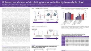 科学海报Unbiased Enrichment of Circulating Tumour Cells Directly from Whole Blood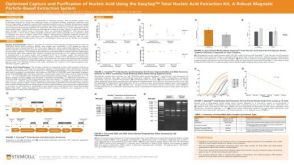


 沪公网安备31010102008431号