若您需要咨询产品或有任何技术问题,请通过官方电话 400 885 9050 或邮箱 info.cn@stemcell.com 与我们联系。

EasySep™人CD45去除试剂盒II

免疫磁珠法去除人CD45+细胞
只有 %1
¥11,820.00

产品号 #(选择产品)

产品号 #17898_C

免疫磁珠法去除人CD45+细胞

产品优势

  • 快捷、操作简单,且无需分离柱
  • CD45+细胞的去除效率高达4 log
  • 分选得到的细胞不带标记

产品组分包括

  • EasySep™人CD45去除试剂盒II(产品号 #17898)
    • EasySep™人CD45去除抗体混合物II,1mL
    • EasySep™ Dextran RapidSpheres™ 磁珠,2x1mL
  • RoboSep™人CD45去除试剂盒II(产品号 #17898RF)
    • EasySep™人CD45去除抗体混合物II,1mL
    • EasySep™ Dextran RapidSpheres™  磁珠,2x1mL
    • RoboSep™ 缓冲液(产品号 #20104)
    • RoboSep™过滤吸头(产品号 #20125)
New look, same high quality and support! You may notice that your instrument or reagent packaging looks slightly different from images displayed on the website, or from previous orders. We are updating our look but rest assured, the products themselves and how you should use them have not changed. Learn more
专为您的实验方案打造的产品
要查看实验方案所需的所有配套产品,请参阅《实验方案与技术文档》

总览

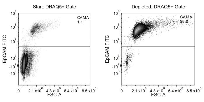
使用EasySep™人CD45去除试剂盒II,可通过免疫磁珠选择高效去除新鲜或冻存人外周血单个核细胞(PBMC)样本中的CD45+细胞。EasySep™技术结合单克隆抗体的特异性和免分离柱系统的简便性,已在发表的研究中广泛应用超过20年。

此简单优化的EasySep™流程包含用可以识别CD45的抗体复合物和磁珠标记细胞,之后标记的细胞通过EasySep™磁铁分离,最后只需通过简单倾倒去除未标记细胞即可。CD45细胞保留在管中。分选后的细胞可立即用于下游应用,例如流式细胞术、培养或DNA/RNA 提取。CD45抗原在除红细胞和血小板外的所有血源细胞上表达。

该产品可替代EasySep™人CD45去除试剂盒 (产品号 #18259) 以进行更快的细胞分选。

了解更多关于免疫磁珠EasySep™技术的工作原理,或如何通过RoboSep™实现免疫磁珠细胞分选全自动化。探索更多产品优化您的实验流程优化,包括培养基、添加剂、抗体等。

磁极兼容性
• EasySep™磁极(产品号 #18000)
• “The Big Easy” EasySep™磁极(产品号 #18001)
• RoboSep™-S(产品号 #21000)
 
分类
细胞分选试剂盒
 
细胞类型
癌细胞及细胞系
 
种属

 
样本来源
PBMC
 
分选方法
去除
 
应用
细胞分选
 
品牌
EasySep,RoboSep
 
研究领域
癌症,免疫,干细胞生物学
 

实验数据

Typical EasySep™ Human CD45 Depletion Profile

Figure 1. Typical EasySep™ Human CD45 Depletion Profile

In the example above, CAMA cells were seeded into PBMCs at a starting frequency of 1.1% (98.9% CD45+; gated on DRAQ5™ for nucleated cells). The CAMA cell (EpCAM+) content of the depleted fraction is 98%, which is a 4.0 log depletion of CD45+ cells.
NOTE: EpCAM is an antibody against an epithelial cell surface antigen expressed on CAMA cells.

产品说明书及文档

请在《产品说明书》中查找相关支持信息和使用说明,或浏览下方更多实验方案。

Document Type
Product Name
Catalog #
Lot #
Language
Catalog #
17898RF
Lot #
All
Language
English
Catalog #
17898
Lot #
All
Language
English
Document Type
Safety Data Sheet 1
Catalog #
17898RF
Lot #
All
Language
English
Document Type
Safety Data Sheet 2
Catalog #
17898RF
Lot #
All
Language
English
Document Type
Safety Data Sheet 3
Catalog #
17898RF
Lot #
All
Language
English
Document Type
Safety Data Sheet 1
Catalog #
17898
Lot #
All
Language
English
Document Type
Safety Data Sheet 2
Catalog #
17898
Lot #
All
Language
English

相关材料与文献

技术资料 (5)

文献 (7)

Axillary adipose tissue–derived lymphatic endothelial cells exhibit distinct transcriptomic signatures reflecting lymphatic invasion status in breast cancer Breast Cancer Research : BCR 2025 Jun

Abstract

BackgroundLymphatics provide a route for breast cancer cells to metastasize. Lymphatic endothelial cells (LECs), which form the structure of lymphatic vessels, play a key role in this process. Although LECs are pivotal in cancer progression, studies often rely on commercially available cell lines that may not accurately reflect the tumor microenvironment. Therefore, there is a pressing need to directly study patient-derived LECs to better understand their role in breast cancer.MethodsThis study developed a method to isolate and characterize LECs directly from human breast-to-axilla adipose tissue. We used magnetic cell separation to remove CD45 + leukocytes and fluorescence-activated cell sorting to isolate cells expressing CD31 and podoplanin. Isolated cells were cultured under conditions promoting endothelial cell growth and were characterized through various assays assessing proliferation, tube formation, and gene expression patterns.ResultsThe sorted CD31 + /PDPN + /CD45 − cell populations exhibited marked increases in proliferation upon VEGF-C stimulation and formed tubule structures on BME-coated dishes, confirming their LEC properties. Notably, isolated LECs showed distinct gene expression patterns depending on the presence of lymph node metastasis and lymphatic invasion.ConclusionsThe ability to isolate and characterize patient-derived LECs from mammary adipose tissue offers new insights into the cellular mechanisms underlying breast cancer metastasis. Significant gene expression variability related to disease state highlights the potential of these cells as biomarkers and therapeutic targets. This study emphasizes the importance of using patient-derived cells to accurately assess the tumor microenvironment, potentially leading to more personalized therapeutic approaches.Supplementary InformationThe online version contains supplementary material available at 10.1186/s13058-025-02067-w.
A FACS-based novel isolation technique identifies heterogeneous CTCs in oral squamous cell carcinoma Frontiers in Oncology 2024 Feb

Abstract

PurposeIsolating circulating tumour cells (CTCs) from the blood is challenging due to their low abundance and heterogeneity. Limitations of conventional CTC detection methods highlight the need for improved strategies to detect and isolate CTCs. Currently, the Food and Drug Administration (FDA)-approved CellSearch™ and other RUO techniques are not available in India. Therefore, we wanted to develop a flexible CTC detection/isolation technique that addresses the limitation(s) of currently available techniques and is suitable for various downstream applications.MethodsWe developed a novel, efficient, user-friendly CTC isolation strategy combining density gradient centrifugation and immuno-magnetic hematogenous cell depletion with fluorescence-activated cell sorting (FACS)-based positive selection using multiple CTC-specific cell-surface markers. For FACS, a stringent gating strategy was optimised to exclude debris and doublets by side scatter/forward scatter (SSC/FSC) discriminator, remove dead cells by 4′,6-diamidino-2-phenylindole (DAPI) staining, and eliminate non-specific fluorescence using a “dump” channel. APC-labelled anti-CD45mAB was used to gate remaining hematogenous cells, while multiple epithelial markers (EpCAM, EGFR, and Pan-Cytokeratin) and an epithelial–mesenchymal transition (EMT) marker (Vimentin) labelled with fluorescein isothiocyanate (FITC) were used to sort cancer cells. The technique was initially developed by spiking Cal 27 cancer cells into the blood of healthy donors and then validated in 95 biopsy-proven oral squamous cell carcinoma (OSCC) patients. CTCs isolated from patients were reconfirmed by Giemsa staining, immuno-staining, and whole transcriptome amplification (WTA), followed by qRT-PCR. In vitro culture and RNA sequencing (RNA-Seq) were also performed to confirm their suitability for various downstream applications.ResultsThe mean detection efficiency for the Cal 27 tongue cancer cells spiked in the whole blood of healthy donors was 32.82% ± 12.71%. While ~75% of our patients (71/95) had detectable CTCs, the CTC positivity was independent of the TNM staging. The isolated potential cancer cells from OSCC patients were heterogeneous in size. They expressed different CTC-specific markers in various combinations as identified by qRT-PCR after WTA in different patients. Isolated CTCs were also found to be suitable for downstream applications like short-term CTC culture and RNA-Seq.ConclusionWe developed a sensitive, specific, flexible, and affordable CTC detection/isolation technique, which is scalable to larger patient cohorts, provides a snapshot of CTC heterogeneity, isolates live CTCs ready for downstream molecular analysis, and, most importantly, is suitable for developing countries.
Pseudo-mutant P53 is a unique phenotype of DNMT3A-mutated pre-leukemia. A. Tuval et al. Haematologica 2022 nov

Abstract

Pre-leukemic clones carrying DNMT3A mutations have a selective advantage and an inherent chemoresistance, however the basis for this phenotype has not been fully elucidated. Mutations affecting the gene TP53 occur in pre-leukemic hematopoietic stem/progenitor cells (preL-HSPC) and lead to chemoresistance. Many of these mutations cause a conformational change and some of them were shown to enhance self-renewal capacity of preL-HSPC. Intriguingly, a misfolded P53 was described in AML blasts that do not harbor mutations in TP53, emphasizing the dynamic equilibrium between wild-type (WT) and pseudo-mutant" conformations of P53. By combining single cell analyses and P53 conformation-specific monoclonal antibodies we studied preL-HSPC from primary human DNMT3A-mutated AML samples. We found that while leukemic blasts express mainly the WT conformation in preL-HSPC the pseudo-mutant conformation is the dominant. HSPC from non-leukemic samples expressed both conformations to a similar extent. In a mouse model we found a small subset of HSPC with a dominant pseudo-mutant P53. This subpopulation was significantly larger among DNMT3AR882H-mutated HSPC suggesting that while a pre-leukemic mutation can predispose for P53 misfolding additional factors are involved as well. Treatment with a short peptide that can shift the dynamic equilibrium favoring the WT conformation of P53 specifically eliminated preL-HSPC that had dysfunctional canonical P53 pathway activity as reflected by single cell RNA sequencing. Our observations shed light upon a possible targetable P53 dysfunction in human preL-HSPC carrying DNMT3A mutations. This opens new avenues for leukemia prevention."

更多信息

更多信息
物种 人类
Magnet Compatibility • EasySep™ Magnet (Catalog #18000) • “The Big Easy” EasySep™ Magnet (Catalog #18001) • RoboSep™-S (Catalog #21000)
样本来源 PBMC
Selection Method Depletion
标记抗体
质量保证:

产品仅供研究使用,不用于针对人或动物的诊断或治疗。 欲获悉更多关于STEMCELL的质控信息,请访问 STEMCELL.CN/COMPLIANCE.
Copyright © 2025 by STEMCELL Technologies. All rights reserved.

在线联系