若您需要咨询产品或有任何技术问题,请通过官方电话 400 885 9050 或邮箱 info.cn@stemcell.com 与我们联系。

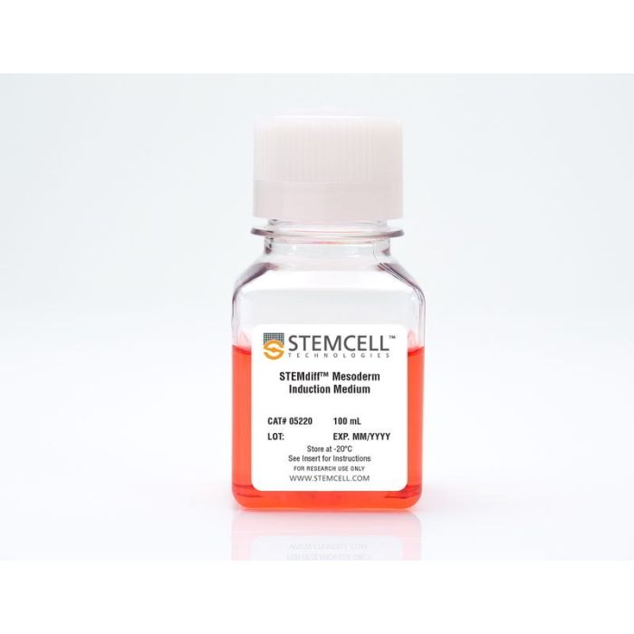
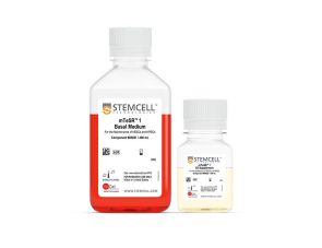
STEMdiff™ 中胚层诱导培养基

用于早期中胚层分化的成分明确、无异种成分的诱导培养基
只有 %1
¥1,774.00

产品号 #(选择产品)

产品号 #05220_C

用于早期中胚层分化的成分明确、无异种成分的诱导培养基

产品优势

  • 成分明确且无异种成分
  • 可在 2–4 天内快速诱导中胚层
  • 高效且可重复地分化多种人ES和iPS细胞系
  • 生成的早期中胚层细胞可进一步分化为多种下游细胞类型
Need a high-quality cell source? Use the hiPSC SCTi003-A (female) or SCTi004-A (male) control lines, manufactured with mTeSR™ Plus.
专为您的实验方案打造的产品
要查看实验方案所需的所有配套产品,请参阅《实验方案与技术文档》

总览

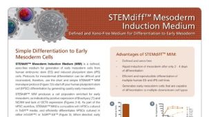
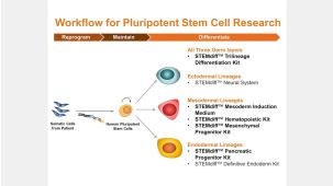
STEMdiff™ 中胚层诱导培养基(MIM)是一种成分明确、无异种成分的培养基,适用于从人胚胎干细胞(ES)和诱导多能干细胞(iPS)中生成早期中胚层细胞。由于中胚层分化的常规方法常常操作复杂且结果不一致,因此推荐使用简短且操作简便的 STEMdiff™ MIM 单层培养方案来诱导人多能干细胞(hPSC)分化。STEMdiff™ MIM 是一种完全培养基,可产生富含早期中胚层的细胞群,其特征为 Brachyury (T) 和 NCAM 标记物的阳性表达。作为人多能干细胞培养流程的一部分,STEMdiff™ MIM 可高效诱导在TeSR™培养基中培养的hPSC进行分化。通过定向诱导,使用 STEMdiff™ MIM 所获得的早期中胚层细胞可进一步分化为多种特化细胞类型,如成骨细胞、软骨细胞、脂肪细胞或内皮细胞。

分类
专用培养基
 
细胞类型
中胚层,PSC衍生,多能干细胞
 
种属

 
应用
细胞培养,分化
 
品牌
STEMdiff
 
研究领域
干细胞生物学
 
制剂类别
无血清,无异源
 

实验数据

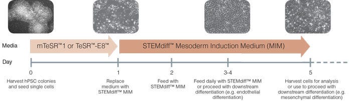
Schematic of Mesoderm Induction Medium Differentiation Timeline

Figure 1. Schematic of Mesoderm Induction Medium Differentiation Timeline

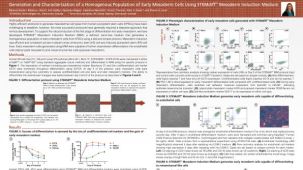
On day 0, hPSC colonies are harvested and seeded as single cells at 5 x 10 4 /cm 2 in mTeSR™1 or TeSR™-E8™ and supplemented with 10 µM Y-27632. TeSR™ medium is replaced on day 1 with STEMdiff™ Mesoderm Induction Medium when cells are at approximately 20 - 50% confluency. Cells are then fed daily and cultured in STEMdiff™ MIM (days 2-4). Cells can be transferred to downstream differentiation conditions between days 3 - 5 or collected on day 5 for analysis.

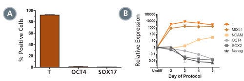
STEMdiff™ MIM Generates a Homogenous Population of T + OCT4 - Early Mesoderm

Figure 2. STEMdiff™ MIM Generates a Homogenous Population of T + OCT4 - Early Mesoderm

(A) Data showing marker expression characteristic of the early mesoderm (positive Brachyury (T) expression and negative OCT4 and SOX 17 expression) on day 5 of the protocol. Data is expressed as a mean percentage of cells expressing each marker ± SD, n = 33 (T, OCT4), n = 5 (SOX17). (B) Expression of undifferentiated cell markers (OCT4, SOX2, NANOG) and early mesoderm markers (T, MIXL1, NCAM), measured by quantitative PCR (qPCR) and normalized to levels in undifferentiated cells, n = 2.

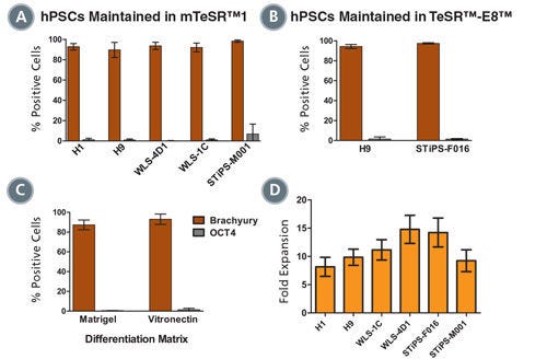
Mesoderm Differentiation and Cell Expansion are Efficient and Comparable Across Multiple hPSC Cell Lines

Figure 3. Mesoderm Differentiation and Cell Expansion are Efficient and Comparable Across Multiple hPSC Cell Lines

Graphs show mesoderm formation in multiple human ES (H1 and H9) and iPS (WLS-4D1, WLS-1C, STiPS-M001 and STiPS-F016) cell lines as measured by expression of Brachyury (T) and absence of OCT4. Cells maintained in (A) mTeSR™1 or (B) TeSR™-E8™ medium were differentiated using STEMdiff™MIM. (A, n = 2 - 10 per cell line, B, n = 3, data are expressed as a mean percentage ± SD) (C) Mesoderm differentiation on Corning® Matrigel® or Vitronectin XF™ is comparable. (n = 5, data are the mean percentage ± SD) (D) Average fold expansion of cells cultured in STEMdiff™MIM, as determined by cell yield / cells seeded. (n = 3 - 13. Error bars indicate SEM)

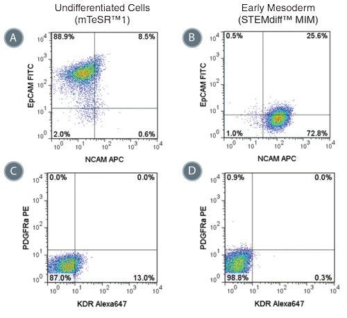
Phenotype of Cells Treated with STEMdiff™ MIM is Consistent with Early Mesoderm

Figure 4. Phenotype of Cells Treated with STEMdiff™ MIM is Consistent with Early Mesoderm

Representative flow cytometry plots showing the switch from (A) EpCAM + NCAM -/low in hPSCs cultured in mTeSR™1 to (B) EpCAM -/low NCAM + expression in STEMdiff™ MIM-treated cells (day 5). EpCAM -/low NCAM + expression is characteristic of the early mesoderm. Expression of PDGFRα and KDR are low in both (C) hPSCs cultured in mTeSR™1 and (D) early mesoderm cells derived with STEMdiff™ MIM.

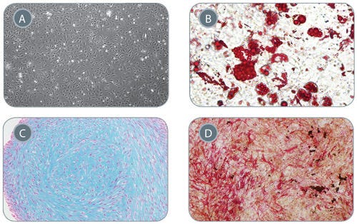
Mesenchymal Stem Cells Derived from Early Mesoderm Cells Can Be Further Differentiated in In Vitro Assays

Figure 5. Mesenchymal Stem Cells Derived from Early Mesoderm Cells Can Be Further Differentiated in In Vitro Assays

(A) Early mesoderm cells generated with the 5-day STEMdiff™ MIM protocol and subsequently cultured with MesenCult™-ACF develop mesenchymal stem cell (MSC)-like morphology, 40X magnifi cation. MSC-like cells can subsequently differentiate into (B) adipocytes (Oil Red O staining), 200x magnification, (C) chondrocytes (Alcian Blue staining), 100X magnification, and (D) osteogenic cells (Fast Red and Silver Nitrate staining), 40X magnification.

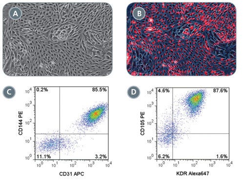
Robust Endothelial Differentiation of STEMdiff™ MIM-Generated Early Mesoderm Cells

Figure 6. Robust Endothelial Differentiation of STEMdiff™ MIM-Generated Early Mesoderm Cells

On day 3 of the STEMdiff™ MIM protocol, early mesoderm cells were switched to a downstream endothelial differentiation protocol based on Tan et al. (A) Differentiated cells display characteristic endothelial cell morphology and (B) are able to uptake Dil-Ac-LDL (red). Representative flow cytometry plots showing (C) 85.5% CD144 + CD31 + and (D) 87.6% CD105 + KDR + expression in differentiated endothelial cells.

产品说明书及文档

请在《产品说明书》中查找相关支持信息和使用说明,或浏览下方更多实验方案。

Document Type
Product Name
Catalog #
Lot #
Language
Catalog #
05221, 05220
Lot #
All
Language
English
Document Type
Safety Data Sheet
Catalog #
05221, 05220
Lot #
All
Language
English

应用领域

本产品专为以下研究领域设计,适用于工作流程中的高亮阶段。探索这些工作流程,了解更多我们为各研究领域提供的其他配套产品。

相关材料与文献

技术资料 (6)

文献 (5)

Highly efficient generation of self-renewing trophoblast from human pluripotent stem cells J. Slamecka et al. iScience 2024 Sep

Abstract

Human pluripotent stem cells (hPSCs) represent a powerful model system to study early developmental processes. However,lineage specification into trophectoderm (TE) and trophoblast (TB) differentiation remains poorly understood,and access to well-characterized placental cells for biomedical research is limited,largely depending on fetal tissues or cancer cell lines. Here,we developed novel strategies enabling highly efficient TE specification that generates cytotrophoblast (CTB) and multinucleated syncytiotrophoblast (STB),followed by the establishment of trophoblast stem cells (TSCs) capable of differentiating into extravillous trophoblast (EVT) and STB after long-term expansion. We confirmed stepwise and controlled induction of lineage- and cell-type-specific genes consistent with developmental biology principles and benchmarked typical features of placental cells using morphological,biochemical,genomics,epigenomics,and single-cell analyses. Charting a well-defined roadmap from hPSCs to distinct placental phenotypes provides invaluable opportunities for studying early human development,infertility,and pregnancy-associated diseases. Subject areas: Natural sciences,Biological sciences,Cell biology,Stem cells research
Cell type- and factor-specific nonsense-mediated RNA decay K. Tan et al. Nucleic Acids Research 2025 May

Abstract

Nonsense-mediated RNA decay (NMD) is a highly conserved RNA turnover pathway that influences several biological processes. Specific features in messenger RNAs (mRNAs) have been found to trigger decay by NMD,leading to the assumption that NMD sensitivity is an intrinsic quality of a given transcript. Here,we provide evidence that,instead,an overriding factor dictating NMD sensitivity is the cell environment. Using several genome-wide techniques to detect NMD-target mRNAs,we find that hundreds of mRNAs are sensitized to NMD as human embryonic stem cells progress to form neural progenitor cells. Another class of mRNAs escape from NMD during this developmental progression. We show that the differential sensitivity to NMD extends to in vivo scenarios,and that the RNA-binding protein,HNRNPL,has a role in cell type-specific NMD. We also addressed another issue in the field—whether NMD factors are core or branch-specific in their action. Surprisingly,we found that UPF3B,an NMD factor critical for the nervous system,shares only 30% of NMD-target transcripts with the core NMD factor UPF2. Together,our findings have implications for how NMD is defined and measured,how NMD acts in different biological contexts,and how different NMD branches influence human diseases.
Endothelial TREM-1 mediates sepsis-induced blood?brain barrier disruption and cognitive impairment via the PI3K/Akt pathway Journal of Neuroinflammation 2025 May

Abstract

The blood?brain barrier (BBB) is a critical selective interface between the central nervous system (CNS) and the blood circulation. BBB dysfunction plays an important role in the neurological damage caused by sepsis. However,the mechanisms underlying the disruption of the BBB during sepsis remain unclear. We established a human induced pluripotent stem cell (iPSC)-derived BBB model and reported that treating with sepsis patient serum leads to structural and functional disruption of the BBB. In a cecal ligation and puncture (CLP)-induced mouse model of sepsis,we also observed disruption of the BBB,inflammation in the brain,and impairments in cognition. In both models,we found that the expression of TREM-1 was significantly increased in endothelial cells. TREM-1 knockout specifically in endothelial cells alleviated BBB dysfunction and cognitive impairments. Further study revealed that TREM-1 affects the expression of genes involved in the PI3K/Akt signaling pathway. The protective effects of TREM-1 inhibition on the BBB and cognition were abrogated by PI3K inhibitors. Our findings suggest that endothelial TREM-1 induces sepsis-induced BBB disruption and cognitive impairments via the PI3K/Akt signaling pathway. Targeting endothelial TREM-1 or the PI3K/Akt signaling pathway may be a promising strategy to maintain BBB integrity and improve cognitive function in sepsis patients.Supplementary InformationThe online version contains supplementary material available at 10.1186/s12974-025-03469-5.

更多信息

更多信息
物种
配方 无血清
质量保证:

产品仅供研究使用,不用于针对人或动物的诊断或治疗。 欲获悉更多关于STEMCELL的质控信息,请访问 STEMCELL.CN/COMPLIANCE.
Copyright © 2026 by STEMCELL Technologies. All rights reserved.

在线联系