
 EasySep™小鼠TIL(CD45)正选试剂盒


技术资料
-

-
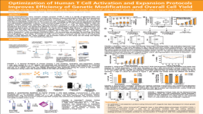 科学海报Optimization of Human T Cell Activation and Expansion Protocols Improves Efficiency of Genetic Modification and Overall Cell Yield
Conference:
AAI 2019
发布日期: 02/14/2020 -
-
 实验方案Enzyme-Free Passaging of Human Pluripotent Stem Cells Using ReLeSR™
研究方向:
干细胞生物学
发布日期: 02/10/2020 -

-
-

-
-
 实验方案How to Process a Leukopak for Downstream Cell Isolation
研究方向:
免疫学,药物发现和毒性检测,传染病
发布日期: 01/21/2020


 沪公网安备31010102008431号