Jason Spence
Developing New Organoid Systems and the Potential Impact of the Technology
研究方向:
呼吸系统研究
Speen AM et al. ( 2016)
Journal of Biological Chemistry 291 48 25192--25206
Ozone-derived oxysterols affect liver X receptor (LXR) signaling: A potential role for lipid-protein adducts
When inhaled,ozone (O3) interacts with cholesterols of airway epithelial cell membranes or the lung lining fluid,generating chemically reactive oxysterols. The mechanism by which O3-derived oxysterols affect molecular function is unknown. Our data show that in vitro exposure of human bronchial epithelial cells to O3 results in the formation of oxysterols,epoxycholesterol-α and β (α-EpCh,β-EpCh) and Secosterol A and B (Seco A,SecoB),in cell lysates and apical washes. Similarly,bronchoalveolar lavage fluid obtained from human volunteers exposed to O3 contained elevated levels of these oxysterol species. As expected,O3-derived oxysterols have a pro-inflammatory effect and increase NF-κB activity. Interestingly,expression of the cholesterol efflux pump ATP-binding cassette transporter 1 (ABCA1),which is regulated by activation of the liver X receptor (LXR),was suppressed in epithelial cells exposed to O3. Additionally,exposure of LXR knockout mice to O3 enhanced pro-inflammatory cytokine production in the lung,suggesting LXR inhibits O3-induced inflammation. Using alkynyl surrogates of O3-derived oxysterols,our data demonstrate adduction of LXR with Seco A. Similarly,supplementation of epithelial cells with alkynyl-tagged cholesterol followed by O3 exposure causes observable lipid-LXR adduct formation. Experiments using Seco A and the LXR agonist T0901317 (T09) showed reduced expression of ABCA1 as compared to stimulation with T09 alone,indicating that Seco A-LXR protein adduct formation inhibits LXR activation by traditional agonists. Overall,these data demonstrate that O3-derived oxysterols have pro-inflammatory functions and form lipid-protein adducts with LXR,thus leading to suppressed cholesterol regulatory gene expression and providing a biochemical mechanism mediating O3-derived formation of oxidized lipids in the airways and subsequent adverse health effects.
View Publication
产品号#:
05001
05021
05022
产品名:
PneumaCult™-ALI 培养基
PneumaCult™-ALI 培养基含12 mm Transwell®插件
PneumaCult™-ALI 培养基含6.5 mm Transwell®插件
Wu Q et al. (NOV 2017)
American journal of physiology. Lung cellular and molecular physiology 30-Nov ajplung003242017
Over-production of growth differentiation factor 15 (GDF15) promotes human rhinovirus infection and virus-induced inflammation in the lung.
Human rhinovirus (HRV) is the most common virus contributing to acute exacerbations of chronic obstructive pulmonary disease (COPD) nearly year-round,but the mechanisms have not been well elucidated. Recent clinical studies suggest that high levels of growth differentiation factor 15 (GDF15) protein in the blood are associated with an increased yearly rate of all-cause COPD exacerbations. Therefore,in the current study,we investigated whether GDF15 promotes HRV infection and virus-induced lung inflammation. We first examined the role of GDF15 in regulating host defense and HRV-induced inflammation using human GDF15 transgenic mice and cultured human GDF15 transgenic mouse tracheal epithelial cells. Next,we determined the effect of GDF15 on viral replication,antiviral responses,and inflammation in human airway epithelial cells with GDF15 knockdown and HRV infection. Finally,we explored the signaling pathways involved in airway epithelial responses to HRV infection in the context of GDF15. Human GDF15 protein over-expression in mice led to exaggerated inflammatory responses to HRV,increased infectious particle release,and decreased IFN-λ2/3 (IL-28A/B) mRNA expression in the lung. Moreover,GDF15 facilitated HRV replication and inflammation via inhibiting IFN-λ1/IL-29 protein production in human airway epithelial cells. Lastly,Smad1 cooperated with interferon regulatory factor 7 (IRF7) to regulate airway epithelial responses to HRV infection partly via GDF15 signaling. Our results reveal a novel function of GDF15 in promoting lung HRV infection and virus-induced inflammation,which may be a new mechanism for the increased susceptibility and severity of respiratory viral (i.e.,HRV) infection in cigarette smoke-exposed airways with GDF15 over-production.
View Publication
产品号#:
05001
05021
05022
产品名:
PneumaCult™-ALI 培养基
PneumaCult™-ALI 培养基含12 mm Transwell®插件
PneumaCult™-ALI 培养基含6.5 mm Transwell®插件
N. J. Ronaghan et al. ( 2022)
PloS one 17 10 e0276013
M1-like, but not M0- or M2-like, macrophages, reduce RSV infection of primary bronchial epithelial cells in a media-dependent fashion.
Respiratory syncytial virus (RSV) is a common childhood infection that in young infants can progress into severe bronchiolitis and pneumonia. Disease pathogenesis results from both viral mediated and host immune processes of which alveolar macrophages play an important part. Here,we investigated the role of different types of alveolar macrophages on RSV infection using an in vitro co-culture model involving primary tissue-derived human bronchial epithelial cells (HBECs) and human blood monocyte-derived M0-like,M1-like,or M2-like macrophages. It was hypothesized that the in vitro model would recapitulate previous in vivo findings of a protective effect of macrophages against RSV infection. It was found that macrophages maintained their phenotype for the 72-hour co-culture time period and the bronchial epithelial cells were unaffected by the macrophage media. HBEC infection with RSV was decreased by M1-like macrophages but enhanced by M0- or M2-like macrophages. The medium used during the co-culture also impacted the outcome of the infection. This work demonstrates that alveolar macrophage phenotypes may have differential roles during epithelial RSV infection,and demonstrates that an in vitro co-culture model could be used to further investigate the roles of macrophages during bronchial viral infection.
View Publication
Cao X et al. (JAN 2015)
Respiratory research 16 30
Tight junction disruption by cadmium in an in vitro human airway tissue model.
BACKGROUND: The cadmium (Cd) present in air pollutants and cigarette smoke has the potential of causing multiple adverse health outcomes involving damage to pulmonary and cardiovascular tissue. Injury to pulmonary epithelium may include alterations in tight junction (TJ) integrity,resulting in impaired epithelial barrier function and enhanced penetration of chemicals and biomolecules. Herein,we investigated mechanisms involved in the disruption of TJ integrity by Cd exposure using an in vitro human air-liquid-interface (ALI) airway tissue model derived from normal primary human bronchial epithelial cells. METHODS: ALI cultures were exposed to noncytotoxic doses of CdCl2 basolaterally and TJ integrity was measured by Trans-Epithelial Electrical Resistance (TEER) and immunofluorescence staining with TJ markers. PCR array analysis was used to identify genes involved with TJ collapse. To explore the involvement of kinase signaling pathways,cultures were treated with CdCl2 in the presence of kinase inhibitors specific for cellular Src or Protein Kinase C (PKC). RESULTS: Noncytotoxic doses of CdCl2 resulted in the collapse of barrier function,as demonstrated by TEER measurements and Zonula occludens-1 (ZO-1) and occludin staining. CdCl2 exposure altered the expression of several groups of genes encoding proteins involved in TJ homeostasis. In particular,down-regulation of select junction-interacting proteins suggested that a possible mechanism for Cd toxicity involves disruption of the peripheral junctional complexes implicated in connecting membrane-bound TJ components to the actin cytoskeleton. Inhibition of kinase signaling using inhibitors specific for cellular Src or PKC preserved the integrity of TJs,possibly by preventing occludin tyrosine hyperphosphorylation,rather than reversing the down-regulation of the junction-interacting proteins. CONCLUSIONS: Our findings indicate that acute doses of Cd likely disrupt TJ integrity in human ALI airway cultures both through occludin hyperphosphorylation via kinase activation and by direct disruption of the junction-interacting complex.
View Publication

 EasySep™小鼠TIL(CD45)正选试剂盒


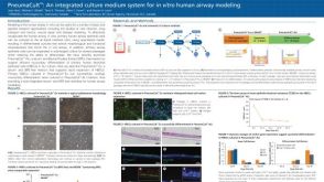 科学海报PneumaCult™: an Integrated Culture Medium System for in Vitro Human Airway Modeling
 专家访谈Jason Spence Developing New Organoid Systems and the Potential Impact of the Technology
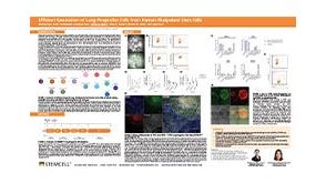 科学海报Efficient Generation of Lung Progenitor Cells From Human Pluripotent Stem Cells
 技术窍门气液界面培养人支气管上皮细胞的关键步骤
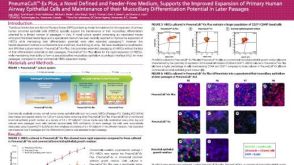 科学海报PneumaCult™-Ex Plus, a Novel Defined and Feeder-Free Medium, Supports the Improved Expansion of Primary Human Airway Epithelial Cells


 沪公网安备31010102008431号