Gomez AM et al. (MAR 2015)
The Journal of Immunology 194 5 2300--8
HIV-1-triggered release of type I IFN by plasmacytoid dendritic cells induces BAFF production in monocytes.
HIV-1 infection leads to numerous B cell abnormalities,including hypergammaglobulinemia,nonspecific B cell activation,nonspecific class switching,increased cell turnover,breakage of tolerance,increased immature/transitional B cells,B cell malignancies,as well as a loss of capacity to generate and maintain memory,all of which contribute to a global impairment of the immune humoral compartment. Several cytokines and soluble factors,which are increased in sera of HIV-1-infected individuals,have been suggested to directly or indirectly contribute to these B cell dysfunctions,and one of these is the B cell-activating factor (BAFF). We report in this study that HIV-1 (X4- and R5-tropic) upregulates BAFF expression and secretion by human monocytes. Moreover,we show that the virus-mediated production of BAFF by monocytes relies on a type I IFN response by a small percentage of plasmacytoid dendritic cells (pDCs) present in the monocyte cultures. HIV-1-induced type I IFN by pDCs triggers BAFF production in both classical and intermediate monocytes,but not in nonclassical monocytes,which nonetheless display a very strong basal BAFF production. We report also that basal BAFF secretion was higher in monocytes obtained from females compared with those from male donors. This study provides a novel mechanistic explanation for the increased BAFF levels observed during HIV-1 infection and highlights the importance of pDC/monocyte crosstalk to drive BAFF secretion.
View Publication
产品号#:
19062
19062RF
19058
19058RF
100-1525
产品名:
EasySep™人浆细胞样DC富集试剂盒
RoboSep™ 人浆细胞样DC富集试剂盒含滤芯吸头
EasySep™人单核细胞富集试剂盒(不去除CD16)
RoboSep™ 人单核细胞富集试剂盒(不去除CD16)含滤芯吸头
EasySep™人单核细胞富集试剂盒(不去除CD16)
Podrazil M et al. (JUL 2015)
Oncotarget 6 20 18192--205
Phase I/II clinical trial of dendritic-cell based immunotherapy (DCVAC/PCa) combined with chemotherapy in patients with metastatic, castration-resistant prostate cancer.
PURPOSE We conducted an open-label,single-arm Phase I/II clinical trial in metastatic CRPC (mCRPC) patients eligible for docetaxel combined with treatment with autologous mature dendritic cells (DCs) pulsed with killed LNCaP prostate cancer cells (DCVAC/PCa). The primary and secondary endpoints were safety and immune responses,respectively. Overall survival (OS),followed as a part of the safety evaluation,was compared to the predicted OS according to the Halabi and MSKCC nomograms. EXPERIMENTAL DESIGN Twenty-five patients with progressive mCRPC were enrolled. Treatment comprised of initial 7 days administration of metronomic cyclophosphamide 50 mg p.o. DCVAC/PCa treatment consisted of a median twelve doses of 1 × 10⁷ dendritic cells per dose injected s.c. (Aldara creme was applied at the site of injection) during a one-year period. The initial 2 doses of DCVAC/PCa were administered at a 2-week interval,followed by the administration of docetaxel (75 mg/m2) and prednisone (5 mg twice daily) given every 3 weeks until toxicity or intolerance was observed. The DCVAC/PCa was then injected every 6 weeks up to the maximum number of doses manufactured from one leukapheresis. RESULTS No serious DCVAC/PCa-related adverse events have been reported. The median OS was 19 months,whereas the predicted median OS was 11.8 months with the Halabi nomogram and 13 months with the MSKCC nomogram. Kaplan-Meier analyses showed that patients had a lower risk of death compared with both MSKCC (Hazard Ratio 0.26,95% CI: 0.13-0.51) and Halabi (Hazard Ratio 0.33,95% CI: 0.17-0.63) predictions. We observed a significant decrease in Tregs in the peripheral blood. The long-term administration of DCVAC/PCa led to the induction and maintenance of PSA specific T cells. We did not identify any immunological parameter that significantly correlated with better OS. CONCLUSIONS In patients with mCRPC,the combined chemoimmunotherapy with DCVAC/PCa and docetaxel was safe and resulted in longer than expected survival. Concomitant chemotherapy did not preclude the induction of specific anti-tumor cytotoxic T cells.
View Publication
产品号#:
07930
07931
07940
07955
07956
07959
07954
100-1061
07952
产品名:
CryoStor® CS10
CryoStor® CS10
CryoStor® CS10
CryoStor® CS10
CryoStor® CS10
CryoStor® CS10
CryoStor® CS10
Rega A et al. (MAR 2013)
Journal of immunology (Baltimore,Md. : 1950) 190 5 2391--402
Plasmacytoid dendritic cells play a key role in tumor progression in lipopolysaccharide-stimulated lung tumor-bearing mice.
The antitumor activity of LPS was first described by Dr. William Coley. However,its role in lung cancer remains unclear. The aim of our study was to elucidate the dose-dependent effects of LPS (0.1-10 μg/mouse) in a mouse model of B16-F10-induced metastatic lung cancer. Lung tumor growth increased at 3 and 7 d after the administration of low-dose LPS (0.1 μg/mouse) compared with control mice. This was associated with an influx of plasmacytoid dendritic cells (pDCs),regulatory T cells,myeloid-derived suppressor cells,and CD8(+) regulatory T cells. In contrast,high-dose LPS (10 μg/mouse) reduced lung tumor burden and was associated with a greater influx of pDCs,as well as a stronger Th1 and Th17 polarization. Depletion of pDCs during low-dose LPS administration resulted in a decreased lung tumor burden. Depletion of pDCs during high-dose LPS treatment resulted in an increased tumor burden. The dichotomy in LPS effects was due to the phenotype of pDCs,which were immunosuppressive after the low-dose LPS,and Th1- and T cytotoxic-polarizing cells after the high-dose LPS. Adoptive transfer of T cells into nude mice demonstrated that CD8(+) T cells were responsible for pDC recruitment following low-dose LPS administration,whereas CD4(+) T cells were required for pDC influx after the high-dose LPS. In conclusion,our data suggest differential effects of low-dose versus high-dose LPS on pDC phenotype and tumor progression or regression in the lungs of mice.
View Publication
产品号#:
19752
19752RF
19753
19753RF
19764
19764RF
产品名:
EasySep™小鼠浆细胞样DC分选试剂盒
RoboSep™ 小鼠浆细胞样DC分选试剂盒
Dannull J et al. (JUL 2013)
The Journal of clinical investigation 123 7 3135--45
Melanoma immunotherapy using mature DCs expressing the constitutive proteasome.
BACKGROUND Many cancers,including melanoma,exclusively express constitutive proteasomes (cPs) and are unable to express immunoproteasomes (iPs). In contrast,mature DCs used for immunotherapy exclusively express iPs. Since proteasomes generate peptides presented by HLA class I molecules,we hypothesized that mature melanoma antigen-loaded DCs engineered to process antigens through cPs would be superior inducers of antimelanoma immunity in vivo. METHODS Subjects with metastatic melanoma were vaccinated with mature DCs transfected with RNAs encoding melanoma antigens MART1,MAGE-3,gp100,and tyrosinase. These DCs were derived from monocytes that were untransfected (Arm A; n = 4),transfected with control siRNA (Arm B; n = 3),or transfected with siRNAs targeting the 3 inducible iP subunits (Arm C; n = 5). RESULTS Vaccination stimulated antigen-specific T cell responses in all subjects,which peaked after 3-4 vaccinations,but remained elevated in Arm C subjects. Also in Arm C,circulating melanoma cell levels (as detected by quantitative PCR) fell,and T cell lytic activity against autologous melanoma was induced. In HLA-A2 subjects,CD8 T cells that bound tetramers loaded with cP-derived melanoma antigenic peptides were found in the peripheral blood only in Arm C subjects. Of 2 subjects with active disease (both in Arm C),one had a partial clinical response,while the other,who exhibited diffuse dermal and soft tissue metastases,had a complete response. CONCLUSION These results suggest that the efficacy of melanoma DC-based immunotherapy is enhanced when tumor antigen-loaded DCs used for vaccination express cPs. TRIAL REGISTRATION Clinicaltrials.gov NCT00672542. FUNDING Duke Clinical Research Institute/Duke Translational Medicine Institute,Duke Melanoma Consortium,and Duke University Department of Surgery.
View Publication
Distinct signals control the hematopoiesis of lymphoid-related dendritic cells.
The molecular and cellular requirements for the development of different populations of human dendritic cells (DC) were studied. Conditions were defined that support DC production from lymphoid progenitors but that fail to induce DC formation from peripheral monocytes. The production of these lymphoid-related DC was severely blocked when hematopoietic progenitors overexpressed Ik7,a mutant dominant-negative Ikaros protein. In contrast,Ik7 did not block the formation of DC in conditions supporting the development of monocyte-derived DC. Furthermore,Ik7 did not block the formation of monocyte/macrophages and enhanced granulopoiesis. One of the molecular mechanisms mediated by Ik7 appears to be down-regulation of the flt3-receptor mRNA. Thus,distinct signals control the formation of DC demonstrating that some aspects of DC diversity are determined in part by distinct molecular cues at the hematopoietic level. (Blood. 2000;95:128-137)
View Publication
产品号#:
04431
产品名:
MethoCult™ H4431
Dadaglio G et al. (MAR 2002)
Journal of immunology (Baltimore,Md. : 1950) 168 5 2219--24
Efficient in vivo priming of specific cytotoxic T cell responses by neonatal dendritic cells.
In early life,a high susceptibility to infectious diseases as well as a poor capacity to respond to vaccines are generally observed as compared with observations in adults. The mechanisms underlying immune immaturity have not been fully elucidated and could be due to the immaturity of the T/B cell responses and/or to a defect in the nature and quality of Ag presentation by the APC. This prompted us to phenotypically and functionally characterize early life murine dendritic cells (DC) purified from spleens of 7-day-old mice. We showed that neonatal CD11c(+) DC express levels of costimulatory molecules and MHC molecules similar to those of adult DC and are able to fully maturate after LPS activation. Furthermore,we demonstrated that neonatal DC can efficiently take up,process,and present Ag to T cells in vitro and induce specific CTL responses in vivo. Although a reduced number of these cells was observed in the spleen of neonatal mice as compared with adults,this study clearly shows that neonatal DC have full functional capacity and may well prime Ag-specific naive T cells in vivo.
View Publication

 EasySep™小鼠TIL(CD45)正选试剂盒


 技术窍门组织解离方法指南

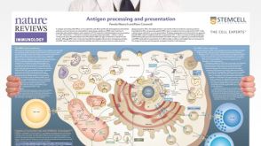 挂图Antigen Processing and Presentation Overview of the mechanisms by which antigens are processed and presented to T cells

 沪公网安备31010102008431号