
 EasySep™小鼠TIL(CD45)正选试剂盒


技术资料
-

-
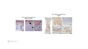
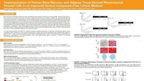 科学海报Characterization of Human Bone Marrow- and Adipose Tissue-Derived Mesenchymal Stromal Cells in an Improved Animal Component-Free Culture Medium
Conference:
ISSCR 2019
发布日期: 11/12/2019 -

-
-
-
-
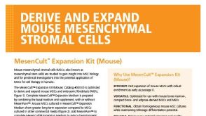
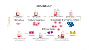
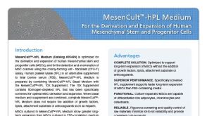 产品手册MesenCult™-hPL Medium For the Derivation and Expansion of Human Mesenchymal Stem and Progenitor Cells
品牌:
MesenCult
发布日期: 07/01/2016

 沪公网安备31010102008431号