Ohoka Y et al. (JAN 2011)
Journal of immunology (Baltimore,Md. : 1950) 186 2 733--44
Retinoic acid-induced CCR9 expression requires transient TCR stimulation and cooperativity between NFATc2 and the retinoic acid receptor/retinoid X receptor complex.
Retinoic acid (RA) imprints gut-homing specificity on T cells upon activation by inducing the expression of chemokine receptor CCR9 and integrin α4β7. CCR9 expression seemed to be more highly dependent on RA than was the α4β7 expression,but its molecular mechanism remained unclear. In this article,we show that NFAT isoforms NFATc1 and NFATc2 directly interact with RA receptor (RAR) and retinoid X receptor (RXR) but play differential roles in RA-induced CCR9 expression on murine naive CD4(+) T cells. TCR stimulation for 6-24 h was required for the acquisition of responsiveness to RA and induced activation of NFATc1 and NFATc2. However,RA failed to induce CCR9 expression as long as TCR stimulation continued. After terminating TCR stimulation or adding cyclosporin A to the culture,Ccr9 gene transcription was induced,accompanied by inactivation of NFATc1 and sustained activation of NFATc2. Reporter and DNA-affinity precipitation assays demonstrated that the binding of NFATc2 to two NFAT-binding sites and that of the RAR/RXR complex to an RA response element half-site in the 5'-flanking region of the mouse Ccr9 gene were critical for RA-induced promoter activity. NFATc2 directly bound to RARα and RXRα,and it enhanced the binding of RARα to the RA response element half-site. NFATc1 also bound to the NFAT-binding sites and directly to RARα and RXRα,but it inhibited the NFATc2-dependent promoter activity. These results suggest that the cooperativity between NFATc2 and the RAR/RXR complex is essential for CCR9 expression on T cells and that NFATc1 interferes with the action of NFATc2.
View Publication
E. Giuliani et al. (mar 2019)
Scientific reports 9 1 4373
Hexamethylene bisacetamide impairs NK cell-mediated clearance of acute T lymphoblastic leukemia cells and HIV-1-infected T cells that exit viral latency.
The hexamethylene bisacetamide (HMBA) anticancer drug was dismissed due to limited efficacy in leukemic patients but it may re-enter into the clinics in HIV-1 eradication strategies because of its recently disclosed capacity to reactivate latent virus. Here,we investigated the impact of HMBA on the cytotoxicity of natural killer (NK) cells against acute T lymphoblastic leukemia (T-ALL) cells or HIV-1-infected T cells that exit from latency. We show that in T-ALL cells HMBA upmodulated MICB and ULBP2 ligands for the NKG2D activating receptor. In a primary CD4+ T cell-based latency model,HMBA did not reactivate HIV-1,yet enhanced ULBP2 expression on cells harboring virus reactivated by prostratin (PRO). However,HMBA reduced the expression of NKG2D and its DAP10 adaptor in NK cells,hence impairing NKG2D-mediated cytotoxicity and DAP10-dependent response to IL-15 stimulation. Alongside,HMBA dampened killing of T-ALL targets by IL-15-activated NK cells and impaired NK cell-mediated clearance of PRO-reactivated HIV-1+ cells. Overall,our results demonstrate a dominant detrimental effect of HMBA on the NKG2D pathway that crucially controls NK cell-mediated killing of tumors and virus-infected cells,providing one possible explanation for poor clinical outcome in HMBA-treated cancer patients and raising concerns for future therapeutic application of this drug.
View Publication
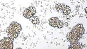
产品号#:
19052
19052RF
产品名:
EasySep™人CD4+ T细胞富集试剂盒
RoboSep™ 人CD4+ T细胞富集试剂盒含滤芯吸头
D. M. Previte et al. (apr 2019)
Cell reports 27 1 129--141.e4
Lymphocyte Activation Gene-3 Maintains Mitochondrial and Metabolic Quiescence in Naive CD4+ T Cells.
Lymphocyte activation gene-3 (LAG-3) is an inhibitory receptor expressed by CD4+ T cells and tempers their homeostatic expansion. Because CD4+ T cell proliferation is tightly coupled to bioenergetics,we investigate the role of LAG-3 in modulating naive CD4+ T cell metabolism. LAG-3 deficiency enhances the metabolic profile of naive CD4+ T cells by elevating levels of mitochondrial biogenesis. In vivo,LAG-3 blockade partially restores expansion and the metabolic phenotype of wild-type CD4+ T cells to levels of Lag3-/- CD4+ T cells,solidifying that LAG-3 controls these processes. Lag3-/- CD4+ T cells also demonstrate greater signal transducer and activator of transcription 5 (STAT5) activation,enabling resistance to interleukin-7 (IL-7) deprivation. These results implicate this pathway as a target of LAG-3-mediated inhibition. Additionally,enhancement of STAT5 activation,as a result of LAG-3 deficiency,contributes to greater activation potential in these cells. These results identify an additional mode of regulation elicited by LAG-3 in controlling CD4+ T cell responses.
View Publication

 EasySep™小鼠TIL(CD45)正选试剂盒


 沪公网安备31010102008431号