Interleukins 7 and 15 Maintain Human T Cell Proliferative Capacity through STAT5 Signaling.
T lymphocytes require signals from self-peptides and cytokines,most notably interleukins 7 and 15 (IL-7,IL-15),for survival. While mouse T cells die rapidly if IL-7 or IL-15 is withdrawn,human T cells can survive prolonged withdrawal of IL-7 and IL-15. Here we show that IL-7 and IL-15 are required to maintain human T cell proliferative capacity through the STAT5 signaling pathway. T cells from humanized mice proliferate better if stimulated in the presence of human IL-7 or IL-15 or if T cells are exposed to human IL-7 or IL-15 in mice. Freshly isolated T cells from human peripheral blood lose proliferative capacity if cultured for 24 hours in the absence of IL-7 or IL-15. We further show that phosphorylation of STAT5 correlates with proliferation and inhibition of STAT5 reduces proliferation. These results reveal a novel role of IL-7 and IL-15 in maintaining human T cell function,provide an explanation for T cell dysfunction in humanized mice,and have significant implications for in vitro studies with human T cells.
View Publication
Richie Ehrlich LI et al. (MAR 2011)
Blood 117 9 2618--24
In vitro assays misrepresent in vivo lineage potentials of murine lymphoid progenitors.
The identity of T-cell progenitors that seed the thymus has remained controversial,largely because many studies differ over whether these progenitors retain myeloid potential. Contradictory reports diverge in their use of various in vitro and in vivo assays. To consolidate these discordant findings,we compared the myeloid potential of 2 putative thymus seeding populations,common lymphoid progenitors (CLPs) and multipotent progenitors (MPPs),and the earliest intrathymic progenitor (DN1),using 2 in vitro assays and in vivo readouts. These assays gave contradictory results: CLP and DN1 displayed surprisingly robust myeloid potential on OP9-DL1 in vitro stromal cocultures but displayed little myeloid potential in vivo,as well as in methylcellulose cultures. MPP,on the other hand,displayed robust myeloid potential in all settings. We conclude that stromal cocultures reveal cryptic,but nonphysiologic,myeloid potentials of lymphoid progenitors,providing an explanation for contradictory findings in the field and underscoring the importance of using in vivo assays for the determination of physiologic lineage potentials.
View Publication
产品号#:
03231
产品名:
MethoCult™ M3231
Serr I et al. (MAR 2016)
Nature Communications 7 10991
Type 1 diabetes vaccine candidates promote human Foxp3(+)Treg induction in humanized mice.
Immune tolerance is executed partly by Foxp3(+)regulatory T (Treg) cells,which suppress autoreactive T cells. In autoimmune type 1 diabetes (T1D) impaired tolerance promotes destruction of insulin-producing β-cells. The development of autoantigen-specific vaccination strategies for Foxp3(+)Treg-induction and prevention of islet autoimmunity in patients is still in its infancy. Here,using human haematopoietic stem cell-engrafted NSG-HLA-DQ8 transgenic mice,we provide direct evidence for human autoantigen-specific Foxp3(+)Treg-induction in vivo. We identify HLA-DQ8-restricted insulin-specific CD4(+)T cells and demonstrate efficient human insulin-specific Foxp3(+)Treg-induction upon subimmunogenic vaccination with strong agonistic insulin mimetopes in vivo. Induced human Tregs are stable,show increased expression of Treg signature genes such as Foxp3,CTLA4,IL-2Rα and TIGIT and can efficiently suppress effector T cells. Such Foxp3(+)Treg-induction does not trigger any effector T cells. These T1D vaccine candidates could therefore represent an expedient improvement in the challenge to induce human Foxp3(+)Tregs and to develop novel precision medicines for prevention of islet autoimmunity in children at risk of T1D.
View Publication
产品号#:
17952
17952RF
100-0696
产品名:
EasySep™人CD4+ T细胞分选试剂盒
RoboSep™ 人CD4+ T细胞分选试剂盒
EasySep™人CD4+ T细胞分离试剂盒
Flach A-C et al. (MAR 2016)
Proceedings of the National Academy of Sciences of the United States of America 113 12 3323--8
Autoantibody-boosted T-cell reactivation in the target organ triggers manifestation of autoimmune CNS disease.
Multiple sclerosis (MS) is caused by T cells that are reactive for brain antigens. In experimental autoimmune encephalomyelitis,the animal model for MS,myelin-reactive T cells initiate the autoimmune process when entering the nervous tissue and become reactivated upon local encounter of their cognate CNS antigen. Thereby,the strength of the T-cellular reactivation process within the CNS tissue is crucial for the manifestation and the severity of the clinical disease. Recently,B cells were found to participate in the pathogenesis of CNS autoimmunity,with several diverse underlying mechanisms being under discussion. We here report that B cells play an important role in promoting the initiation process of CNS autoimmunity. Myelin-specific antibodies produced by autoreactive B cells after activation in the periphery diffused into the CNS together with the first invading pathogenic T cells. The antibodies accumulated in resident antigen-presenting phagocytes and significantly enhanced the activation of the incoming effector T cells. The ensuing strong blood-brain barrier disruption and immune cell recruitment resulted in rapid manifestation of clinical disease. Therefore,myelin oligodendrocyte glycoprotein (MOG)-specific autoantibodies can initiate disease bouts by cooperating with the autoreactive T cells in helping them to recognize their autoantigen and become efficiently reactivated within the immune-deprived nervous tissue.
View Publication

 EasySep™小鼠TIL(CD45)正选试剂盒


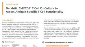 技术公告Dendritic Cell/CD8+ T Cell Co-Culture to Assess Antigen-Specific T Cell Functionality
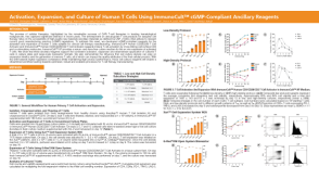 科学海报Activation, Expansion, and Culture of Human T Cells Using ImmunoCult™ cGMP-Compliant Ancillary Reagents

 沪公网安备31010102008431号