Type 1 Interferons Potentiate Human CD8+ T-Cell Cytotoxicity Through a STAT4- and Granzyme B-Dependent Pathway.
Events defining the progression to human type 1 diabetes (T1D) have remained elusive owing to the complex interaction between genetics,the immune system,and the environment. Type 1 interferons (T1-IFN) are known to be a constituent of the autoinflammatory milieu within the pancreas of patients with T1D. However,the capacity of IFNα/β to modulate human activated autoreactive CD8+ T-cell (cytotoxic T lymphocyte) responses within the islets of patients with T1D has not been investigated. Here,we engineer human β-cell-specific cytotoxic T lymphocytes and demonstrate that T1-IFN augments cytotoxicity by inducing rapid phosphorylation of STAT4,resulting in direct binding at the granzyme B promoter within 2 h of exposure. The current findings provide novel insights concerning the regulation of effector function by T1-IFN in human antigen-experienced CD8+ T cells and provide a mechanism by which the presence of T1-IFN potentiates diabetogenicity within the autoimmune islet.
View Publication
产品号#:
15023
15063
产品名:
RosetteSep™人CD8+ T细胞富集抗体混合物
RosetteSep™人CD8+ T细胞富集抗体混合物
Prodeus A et al. (SEP 2017)
JCI insight 2 18
VISTA.COMP - an engineered checkpoint receptor agonist that potently suppresses T cell-mediated immune responses.
V-domain immunoglobulin suppressor of T cell activation (VISTA) is a recently discovered immune checkpoint ligand that functions to suppress T cell activity. The therapeutic potential of activating this immune checkpoint pathway to reduce inflammatory responses remains untapped,largely due to the inability to derive agonists targeting its unknown receptor. A dimeric construct of the IgV domain of VISTA (VISTA-Fc) was shown to suppress the activation of T cells in vitro. However,this effect required its immobilization on a solid surface,suggesting that VISTA-Fc may display limited efficacy as a VISTA-receptor agonist in vivo. Herein,we have designed a stable pentameric VISTA construct (VISTA.COMP) by genetically fusing its IgV domain to the pentamerization domain from the cartilage oligomeric matrix protein (COMP). In contrast to VISTA-Fc,VISTA.COMP does not require immobilization to inhibit the proliferation of CD4+ T cells undergoing polyclonal activation. Furthermore,we show that VISTA.COMP,but not VISTA-Fc,functions as an immunosuppressive agonist in vivo capable of prolonging the survival of skin allografts in a mouse transplant model as well as rescuing mice from acute concanavalin-A-induced hepatitis. Collectively,we believe our data demonstrate that VISTA.COMP is a checkpoint receptor agonist and the first agent to our knowledge targeting the putative VISTA-receptor to suppress T cell-mediated immune responses.
View Publication
产品号#:
19852
19852RF
产品名:
EasySep™小鼠CD4+ T细胞分选试剂盒
RoboSep™ 小鼠CD4+ T细胞分选试剂盒
Halim L et al. (JUL 2017)
Cell reports 20 3 757--770
An Atlas of Human Regulatory T Helper-like Cells Reveals Features of Th2-like Tregs that Support a Tumorigenic Environment.
Regulatory T cells (Tregs) play a pivotal role in maintaining immunological tolerance,but they can also play a detrimental role by preventing antitumor responses. Here,we characterized T helper (Th)-like Treg subsets to further delineate their biological function and tissue distribution,focusing on their possible contribution to disease states. RNA sequencing and functional assays revealed that Th2-like Tregs displayed higher viability and autocrine interleukin-2 (IL-2)-mediated activation than other subsets. Th2-like Tregs were preferentially found in tissues rather than circulation and exhibited the highest migratory capacity toward chemokines enriched at tumor sites. These cellular responses led us to hypothesize that this subset could play a role in maintaining a tumorigenic environment. Concurrently,Th2-like Tregs were enriched specifically in malignant tissues from patients with melanoma and colorectal cancer compared to healthy tissue. Overall,our results suggest that Th2-like Tregs may contribute to a tumorigenic environment due to their increased cell survival,higher migratory capacity,and selective T-effector suppressive ability.
View Publication
D. Duluc et al. ( 2014)
The Journal of Immunology 192 5776-88
Induction and activation of human Th17 by targeting antigens to dendritic cells via dectin-1
Recent compelling evidence indicates that Th17 confer host immunity against a variety of microbes,including extracellular and intracellular pathogens. Therefore,understanding mechanisms for the induction and activation of Ag-specific Th17 is important for the rational design of vaccines against pathogens. To study this,we employed an in vitro system in which influenza hemagglutinin (HA) 1 was delivered to dendritic cells (DCs) via Dectin-1 using anti-human Dectin-1 (hDectin-1)-HA1 recombinant fusion proteins. We found that healthy individuals maintained broad ranges of HA1-specific memory Th17 that were efficiently activated by DCs targeted with anti-hDectin-1-HA1. Nonetheless,these DCs were not able to induce a significant level of HA1-specific Th17 responses even in the presence of the Th17-promoting cytokines IL-1? and IL-6. We further found that the induction of surface IL-1R1 expression by signals via TCRs and common ?-chain receptors was essential for naive CD4(+) T cell differentiation into HA1-specific Th17. This process was dependent on MyD88,but not IL-1R-associated kinase 1/4. Thus,interruptions in STAT3 or MyD88 signaling led to substantially diminished HA1-specific Th17 induction. Taken together,the de novo generation of pathogen-specific human Th17 requires complex,but complementary,actions of multiple signals. Data from this study will help us design a new and effective vaccine strategy that can promote Th17-mediated immunity against microbial pathogens.
View Publication
产品号#:
19052
19052RF
产品名:
EasySep™人CD4+ T细胞富集试剂盒
RoboSep™ 人CD4+ T细胞富集试剂盒含滤芯吸头
Drake A et al. ( 2016)
PloS one 11 11 e0166280
Interleukins 7 and 15 Maintain Human T Cell Proliferative Capacity through STAT5 Signaling.
T lymphocytes require signals from self-peptides and cytokines,most notably interleukins 7 and 15 (IL-7,IL-15),for survival. While mouse T cells die rapidly if IL-7 or IL-15 is withdrawn,human T cells can survive prolonged withdrawal of IL-7 and IL-15. Here we show that IL-7 and IL-15 are required to maintain human T cell proliferative capacity through the STAT5 signaling pathway. T cells from humanized mice proliferate better if stimulated in the presence of human IL-7 or IL-15 or if T cells are exposed to human IL-7 or IL-15 in mice. Freshly isolated T cells from human peripheral blood lose proliferative capacity if cultured for 24 hours in the absence of IL-7 or IL-15. We further show that phosphorylation of STAT5 correlates with proliferation and inhibition of STAT5 reduces proliferation. These results reveal a novel role of IL-7 and IL-15 in maintaining human T cell function,provide an explanation for T cell dysfunction in humanized mice,and have significant implications for in vitro studies with human T cells.
View Publication
S. Gupta et al. ( 2018)
Immunity & ageing : I & A 15 2
Molecular changes associated with increased TNF-?-induced apoptotis in naive (TN) and central memory (TCM) CD8+ T cells in aged humans.
Background Progressive T cell decline in aged humans is associated with a deficiency of naive (TN) and central memory (TCM) T cells. We have previously reported increased tumor necrosis factor-? (TNF-?)-induced apoptosis in TN and TCM T cells in aged humans; however,the molecular basis of increased apoptosis remains to be defined. Since expression of TNF receptors (TNFRs) was reported to be comparable in young and aged,we investigated signaling events downstream of TNFRs to understand the molecular basis of increased TNF-?-induced apoptosis in aged TN and TCM CD8+ cells. Results The expression of TRAF-2 and RIP,phosphorylation of JNK,IKK?/?,and I?B?,and activation of NF-?B activation were significantly decreased in TN and TCM CD8+ cells from aged subjects as compared to young controls. Furthermore,expression of A20,Bcl-xL,cIAP1,and FLIP-L and FLIP-S was significantly decreased in TN and TCM CD8+ cells from aged subjects. Conclusions These data demonstrate that an impaired expression/function of molecules downstream TNFR signaling pathway that confer survival signals contribute to increased apoptosis of TN and TCM CD8+ cells in aged humans.
View Publication

 EasySep™小鼠TIL(CD45)正选试剂盒


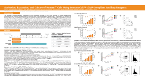 科学海报Activation, Expansion, and Culture of Human T Cells Using ImmunoCult™ cGMP-Compliant Ancillary Reagents


 沪公网安备31010102008431号