Fuller HR et al. (JAN 2015)
Frontiers in cellular neuroscience 9 January 506
Spinal Muscular Atrophy Patient iPSC-Derived Motor Neurons Have Reduced Expression of Proteins Important in Neuronal Development.
Spinal muscular atrophy (SMA) is an inherited neuromuscular disease primarily characterized by degeneration of spinal motor neurons,and caused by reduced levels of the SMN protein. Previous studies to understand the proteomic consequences of reduced SMN have mostly utilized patient fibroblasts and animal models. We have derived human motor neurons from type I SMA and healthy controls by creating their induced pluripotent stem cells (iPSCs). Quantitative mass spectrometry of these cells revealed increased expression of 63 proteins in control motor neurons compared to respective fibroblasts,whereas 30 proteins were increased in SMA motor neurons vs. their fibroblasts. Notably,UBA1 was significantly decreased in SMA motor neurons,supporting evidence for ubiquitin pathway defects. Subcellular distribution of UBA1 was predominantly cytoplasmic in SMA motor neurons in contrast to nuclear in control motor neurons; suggestive of neurodevelopmental abnormalities. Many of the proteins that were decreased in SMA motor neurons,including beta III-tubulin and UCHL1,were associated with neurodevelopment and differentiation. These neuron-specific consequences of SMN depletion were not evident in fibroblasts,highlighting the importance of iPSC technology. The proteomic profiles identified here provide a useful resource to explore the molecular consequences of reduced SMN in motor neurons,and for the identification of novel biomarker and therapeutic targets for SMA.
View Publication
The genetic reprogramming technology allows one to generate pluripotent stem cells for individual patients. These cells,called induced pluripotent stem cells (iPSCs),can be an unlimited source of specialized cell types for the body. Thus,autologous somatic cell replacement therapy becomes possible,as well as the generation of in vitro cell models for studying the mechanisms of disease pathogenesis and drug discovery. Amyotrophic lateral sclerosis (ALS) is an incurable neurodegenerative disorder that leads to a loss of upper and lower motor neurons. About 10% of cases are genetically inherited,and the most common familial form of ALS is associated with mutations in the SOD1 gene. We used the reprogramming technology to generate induced pluripotent stem cells with patients with familial ALS. Patient-specific iPS cells were obtained by both integration and transgene-free delivery methods of reprogramming transcription factors. These iPS cells have the properties of pluripotent cells and are capable of direct differentiation into motor neurons.
View Publication
产品号#:
05850
05857
05870
05875
85850
85857
85870
85875
产品名:
mTeSR™1
mTeSR™1
de Boer AS et al. (AUG 2014)
Science Translational Medicine 6 248 248ra104--248ra104
Genetic validation of a therapeutic target in a mouse model of ALS
AbstractBack to TopbackslashnNeurons produced from stem cells have emerged as a tool to identify new therapeutic targets for neurological diseases such as amyotrophic lateral sclerosis (ALS). However,it remains unclear to what extent these new mechanistic insights will translate to animal models,an important step in the validation of new targets. Previously,we found that glia from mice carrying the SOD1G93A mutation,a model of ALS,were toxic to stem cell–derived human motor neurons. We use pharmacological and genetic approaches to demonstrate that the prostanoid receptor DP1 mediates this glial toxicity. Furthermore,we validate the importance of this mechanism for neural degeneration in vivo. Genetic ablation of DP1 in SOD1G93A mice extended life span,decreased microglial activation,and reduced motor neuron loss. Our findings suggest that blocking DP1 may be a therapeutic strategy in ALS and demonstrate that discoveries from stem cell models of disease can be corroborated in vivo.
View Publication
产品号#:
05850
05857
05870
05875
85850
85857
85870
85875
产品名:
mTeSR™1
mTeSR™1
Musah S et al. (SEP 2014)
Proceedings of the National Academy of Sciences of the United States of America 111 38 13805--10
Substratum-induced differentiation of human pluripotent stem cells reveals the coactivator YAP is a potent regulator of neuronal specification.
Physical stimuli can act in either a synergistic or antagonistic manner to regulate cell fate decisions,but it is less clear whether insoluble signals alone can direct human pluripotent stem (hPS) cell differentiation into specialized cell types. We previously reported that stiff materials promote nuclear localization of the Yes-associated protein (YAP) transcriptional coactivator and support long-term self-renewal of hPS cells. Here,we show that even in the presence of soluble pluripotency factors,compliant substrata inhibit the nuclear localization of YAP and promote highly efficient differentiation of hPS cells into postmitotic neurons. In the absence of neurogenic factors,the effective substrata produce neurons rapidly (2 wk) and more efficiently (textgreater75%) than conventional differentiation methods. The neurons derived from substrate induction express mature markers and possess action potentials. The hPS differentiation observed on compliant surfaces could be recapitulated on stiff surfaces by adding small-molecule inhibitors of F-actin polymerization or by depleting YAP. These studies reveal that the matrix alone can mediate differentiation of hPS cells into a mature cell type,independent of soluble inductive factors. That mechanical cues can override soluble signals suggests that their contributions to early tissue development and lineage commitment are profound.
View Publication
产品号#:
05850
05857
05870
05875
85850
85857
85870
85875
产品名:
mTeSR™1
mTeSR™1
Saporta MA et al. (JAN 2015)
Experimental neurology 263 190--199
Axonal Charcot-Marie-Tooth disease patient-derived motor neurons demonstrate disease-specific phenotypes including abnormal electrophysiological properties
OBJECTIVE Charcot-Marie-Tooth (CMT) disease is a group of inherited peripheral neuropathies associated with mutations or copy number variations in over 70 genes encoding proteins with fundamental roles in the development and function of Schwann cells and peripheral axons. Here,we used iPSC-derived cells to identify common pathophysiological mechanisms in axonal CMT. METHODS iPSC lines from patients with two distinct forms of axonal CMT (CMT2A and CMT2E) were differentiated into spinal cord motor neurons and used to study axonal structure and function and electrophysiological properties in vitro. RESULTS iPSC-derived motor neurons exhibited gene and protein expression,ultrastructural and electrophysiological features of mature primary spinal cord motor neurons. Cytoskeletal abnormalities were found in neurons from a CMT2E (NEFL) patient and corroborated by a mouse model of the same NEFL point mutation. Abnormalities in mitochondrial trafficking were found in neurons derived from this patient,but were only mildly present in neurons from a CMT2A (MFN2) patient. Novel electrophysiological abnormalities,including reduced action potential threshold and abnormal channel current properties were observed in motor neurons derived from both of these patients. INTERPRETATION Human iPSC-derived motor neurons from axonal CMT patients replicated key pathophysiological features observed in other models of MFN2 and NEFL mutations,including abnormal cytoskeletal and mitochondrial dynamics. Electrophysiological abnormalities found in axonal CMT iPSC-derived human motor neurons suggest that these cells are hyperexcitable and have altered sodium and calcium channel kinetics. These findings may provide a new therapeutic target for this group of heterogeneous inherited neuropathies.
View Publication
产品号#:
05850
05857
05870
05875
85850
85857
85870
85875
产品名:
mTeSR™1
mTeSR™1
Schrenk-Siemens K et al. (JAN 2014)
Nature neuroscience 18 1 10--16
PIEZO2 is required for mechanotransduction in human stem cell-derived touch receptors.
Human sensory neurons are inaccessible for functional examination,and thus little is known about the mechanisms mediating touch sensation in humans. Here we demonstrate that the mechanosensitivity of human embryonic stem (hES) cell-derived touch receptors depends on PIEZO2. To recapitulate sensory neuron development in vitro,we established a multistep differentiation protocol and generated sensory neurons via the intermediate production of neural crest cells derived from hES cells or human induced pluripotent stem (hiPS) cells. The generated neurons express a distinct set of touch receptor-specific genes and convert mechanical stimuli into electrical signals,their most salient characteristic in vivo. Strikingly,mechanosensitivity is lost after CRISPR/Cas9-mediated PIEZO2 gene deletion. Our work establishes a model system that resembles human touch receptors,which may facilitate mechanistic analysis of other sensory subtypes and provide insight into developmental programs underlying sensory neuron diversity.
View Publication
产品号#:
05850
05857
05870
05875
85850
85857
85870
85875
产品名:
mTeSR™1
mTeSR™1
Zhao HW et al. (MAR 2015)
Neuroscience 288 187--199
Altered iPSC-derived neurons' sodium channel properties in subjects with Monge's disease
Monge's disease,also known as chronic mountain sickness (CMS),is a disease that potentially threatens more than 140 million highlanders during extended time living at high altitudes (over 2500m). The prevalence of CMS in Andeans is about 15-20%,suggesting that the majority of highlanders (non-CMS) are rather healthy at high altitudes; however,CMS subjects experience severe hypoxemia,erythrocytosis and many neurologic manifestations including migraine,headache,mental fatigue,confusion,and memory loss. The underlying mechanisms of CMS neuropathology are not well understood and no ideal treatment is available to prevent or cure CMS,except for phlebotomy. In the current study,we reprogrammed fibroblast cells from both CMS and non-CMS subjects' skin biopsies into the induced pluripotent stem cells (iPSCs),then differentiated into neurons and compared their neuronal properties. We discovered that CMS neurons were much less excitable (higher rheobase) than non-CMS neurons. This decreased excitability was not caused by differences in passive neuronal properties,but instead by a significantly lowered Na+ channel current density and by a shift of the voltage-conductance curve in the depolarization direction. Our findings provide,for the first time,evidence of a neuronal abnormality in CMS subjects as compared to non-CMS subjects,hoping that such studies can pave the way to a better understanding of the neuropathology in CMS.
View Publication
产品号#:
05850
05857
05870
05875
85850
85857
85870
85875
产品名:
mTeSR™1
mTeSR™1
Pei Y et al. (MAY 2016)
Brain research 1638 Pt A 57--73
Comparative neurotoxicity screening in human iPSC-derived neural stem cells, neurons and astrocytes.
Induced pluripotent stem cells (iPSC) and their differentiated derivatives offer a unique source of human primary cells for toxicity screens. Here,we report on the comparative cytotoxicity of 80 compounds (neurotoxicants,developmental neurotoxicants,and environmental compounds) in iPSC as well as isogenic iPSC-derived neural stem cells (NSC),neurons,and astrocytes. All compounds were tested over a 24-h period at 10 and 100$\$,in duplicate,with cytotoxicity measured using the MTT assay. Of the 80 compounds tested,50 induced significant cytotoxicity in at least one cell type; per cell type,32,38,46,and 41 induced significant cytotoxicity in iPSC,NSC,neurons,and astrocytes,respectively. Four compounds (valinomycin,3,3',5,5'-tetrabromobisphenol,deltamethrin,and triphenyl phosphate) were cytotoxic in all four cell types. Retesting these compounds at 1,10,and 100$\$ using the same exposure protocol yielded consistent results as compared with the primary screen. Using rotenone,we extended the testing to seven additional iPSC lines of both genders; no substantial difference in the extent of cytotoxicity was detected among the cell lines. Finally,the cytotoxicity assay was simplified by measuring luciferase activity using lineage-specific luciferase reporter iPSC lines which were generated from the parental iPSC line. This article is part of a Special Issue entitled SI: PSC and the brain.
View Publication
产品号#:
05850
05857
05870
05875
85850
85857
85870
85875
产品名:
mTeSR™1
mTeSR™1
Sun Y et al. (MAR )
PLOS ONE 3 e0118771
Properties of Neurons Derived from Induced Pluripotent Stem Cells of Gaucher Disease Type 2 Patient Fibroblasts: Potential Role in Neuropathology
Gaucher disease (GD) is caused by insufficient activity of acid $\$-glucosidase (GCase) resulting from mutations in GBA1. To understand the pathogenesis of the neuronopathic GD,induced pluripotent stem cells (iPSCs) were generated from fibroblasts isolated from three GD type 2 (GD2) and 2 unaffected (normal and GD carrier) individuals. The iPSCs were converted to neural precursor cells (NPCs) which were further differentiated into neurons. Parental GD2 fibroblasts as well as iPSCs,NPCs,and neurons had similar degrees of GCase deficiency. Lipid analyses showed increases of glucosylsphingosine and glucosylceramide in the GD2 cells. In addition,GD2 neurons showed increased $\$-synuclein protein compared to control neurons. Whole cell patch-clamping of the GD2 and control iPSCs-derived neurons demonstrated excitation characteristics of neurons,but intriguingly,those from GD2 exhibited consistently less negative resting membrane potentials with various degree of reduction in action potential amplitudes,sodium and potassium currents. Culture of control neurons in the presence of the GCase inhibitor (conduritol B epoxide) recapitulated these findings,providing a functional link between decreased GCase activity in GD and abnormal neuronal electrophysiological properties. To our knowledge,this study is first to report abnormal electrophysiological properties in GD2 iPSC-derived neurons that may underlie the neuropathic phenotype in Gaucher disease.
View Publication
Lippmann ES et al. (APR 2014)
Stem Cells 32 4 1032--1042
Defined human pluripotent stem cell culture enables highly efficient neuroepithelium derivation without small molecule inhibitors.
The embryonic neuroepithelium gives rise to the entire central nervous system in vivo,making it an important tissue for developmental studies and a prospective cell source for regenerative applications. Current protocols for deriving homogenous neuroepithelial cultures from human pluripotent stem cells (hPSCs) consist of either embryoid body-mediated neuralization followed by a manual isolation step or adherent differentiation using small molecule inhibitors. Here,we report that hPSCs maintained under chemically defined,feeder-independent,and xeno-free conditions can be directly differentiated into pure neuroepithelial cultures ([mt]90% Pax6(+)/N-cadherin(+) with widespread rosette formation) within 6 days under adherent conditions,without small molecule inhibitors,and using only minimalistic medium consisting of Dulbecco's modified Eagle's medium/F-12,sodium bicarbonate,selenium,ascorbic acid,transferrin,and insulin (i.e.,E6 medium). Furthermore,we provide evidence that the defined culture conditions enable this high level of neural conversion in contrast to hPSCs maintained on mouse embryonic fibroblasts (MEFs). In addition,hPSCs previously maintained on MEFs could be rapidly converted to a neural compliant state upon transfer to these defined conditions while still maintaining their ability to generate all three germ layers. Overall,this fully defined and scalable protocol should be broadly useful for generating therapeutic neural cells for regenerative applications.
View Publication


 EasySep™小鼠TIL(CD45)正选试剂盒




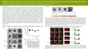 科学海报STEMdiff™ Cerebral Organoid Kit: A New Tool for the Culture of 3D Brain Organoids Derived from hPSCs
 沪公网安备31010102008431号