若您需要咨询产品或有任何技术问题,请通过官方电话 400 885 9050 或邮箱 info.cn@stemcell.com 与我们联系。

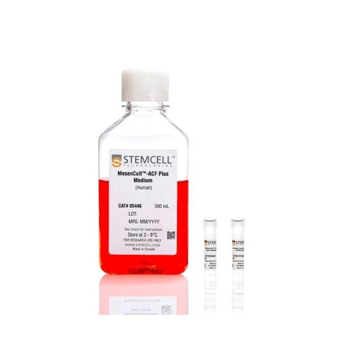
MesenCult™-ACF Plus培养试剂盒

用于人间充质干细胞的无动物成分培养基及贴附基质
只有 %1
¥4,542.00

产品号 #(选择产品)

产品号 #05448

用于人间充质干细胞的无动物成分培养基及贴附基质

产品优势

  • 无动物成分配方提升实验可重复性。
  • 相较于含血清培养基,具备更优异的细胞扩增效果。
  • 培养的MSC在早期和晚期传代中均保持强劲的扩增能力及三系分化潜能。
  • 支持直接从人类原代组织中衍生MSC。

产品组分包括

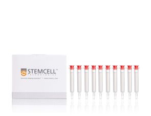
  • MesenCult™ -ACF Plus基础培养基(500 mL)
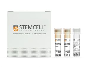
  • MesenCult™-ACF Plus 500X 补充剂(1 mL)
  • 无动物成分细胞贴附基质 (1 mL)
专为您的实验方案打造的产品
要查看实验方案所需的所有配套产品,请参阅《实验方案与技术文档》

总览

使用MesenCult™-ACF Plus培养试剂盒减少人间充质基质细胞(MSC)培养的变异性并提高可重复性。该试剂盒不含动物源性成分(ACF)和细胞外囊泡(EV),并针对多种来源(包括骨髓和脂肪组织)的MSC的无血清诱导和扩增进行了优化。

MesenCult™-ACF Plus支持高效的MSC扩增,同时保持干净、无EV的培养背景。由于该配方不含外源EVs,您可以直接从条件培养基中分离MSC来源的EVs,而无需切换到专门的EV收集培养基。有关使用此系统生成EV的指导说明,请参阅我们详细的EV生成方案。将该试剂盒与EasySep™人细胞外囊泡正选试剂盒</a>配合使用,以实现高效和标准化的EV回收。

与含血清或去除EV的培养基相比,在MesenCult™-ACF Plus中培养的MSC能够更高效地扩增并保持标志性的MSC特征,包括表面标志物表达、强效增殖以及可通过MesenCult™-ACF软骨生成分化试剂盒MesenCult™脂肪生成分化试剂盒(人)MesenCult™成骨分化试剂盒(人)进行评估的三谱系分化潜能。

该试剂盒是更广泛的MesenCult™ MSC细胞培养产品组合的一部分,该产品组合包含用于MSC分化、培养和鉴定的试剂。您还可以使用人iPSC衍生的间充质祖细胞,从经过验证、纯的MSC培养物开始您的实验。

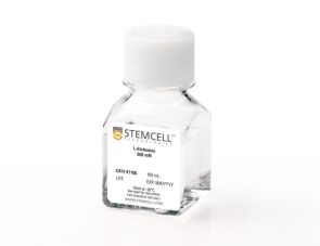
注意:MesenCult™-ACF Plus完全培养基必须添加L-谷氨酰胺。CollPlant是细胞贴附基质中重组人胶原蛋白(rhCollagen)成分的制造商。


分类
专用培养基
 
细胞类型
间充质细胞,PSC衍生,间充质干/祖细胞
 
种属

 
应用
细胞培养,扩增,培养
 
品牌
MesenCult
 
研究领域
药物发现和毒性检测,细胞外囊泡研究,干细胞生物学
 
制剂类别
不含动物成分,无血清
 

实验数据

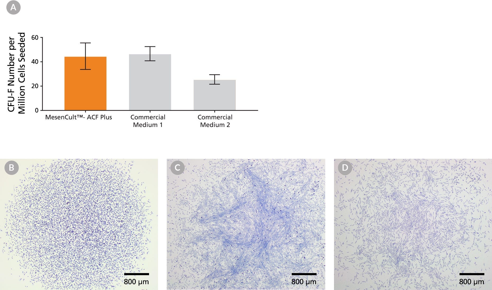
Figure 1. CFU-F Assay of Human BM-Derived MSCs Expanded in MesenCult™-ACF Plus Medium and Commercial Media.
(A) An average of 45 CFU-Fs per million cells were observed when BM mononuclear cells were seeded in MesenCult™-ACF Plus (n = 4). An average of 47 and 25 CFU-Fs per million cells were observed when cells were seeded in Commercial Medium 1 (n = 3) and Medium 2 (n = 4), respectively. Vertical lines indicate Standard Error of Mean (SEM). Representative image of CFU-F colonies expanded in (B) MesenCult™-ACF Plus Medium (9 days of culture), (C) Commercial Medium 1 (10 days of culture) and (D) Commercial Medium 2 (10 days of culture). Commercial Medium 1 and Medium 2 were supplemented with 2.5% human AB serum to derive MSCs from BM, as per their protocols for derivation. No addition of serum is required when using MesenCult™-ACF Plus Medium.

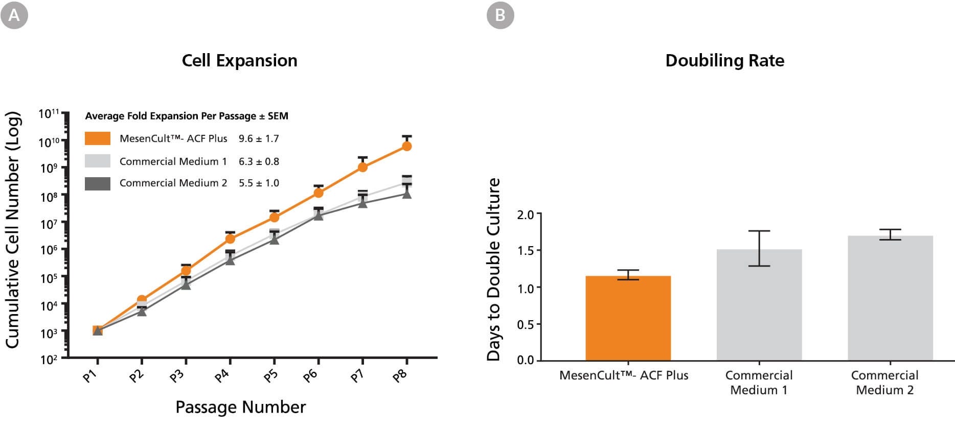
Figure 2. Human BM-Derived MSCs Cultured in MesenCult™-ACF Plus Medium Expand Faster than MSCs Cultured in Commercial Xeno-Free and Serum-Free Media.
(A) A greater number of BM-derived MSCs were generated per passage using MesenCult™-ACF Plus Medium (n=4) compared to Commercial Medium 1 (n=3) and Commercial Medium 2 (n=2). (B) Rates of BM-derived MSC expansion were compared between MesenCult™-ACF Plus Medium, Commercial Medium 1, and Commercial Medium 2. The time required to double the number of MSCs using MesenCult™ -ACF Plus Medium (n=4) was shorter than when MSCs were cultured in Commercial Medium 1 (n=3) and Commercial Medium 2 (n=4). Vertical lines indicate Standard Error of Mean (SEM).

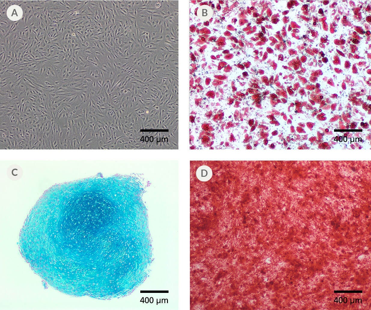
Figure 3. Human BM-Derived MSCs Expanded in MesenCult™-ACF Plus Medium Display Multi-Lineage Differentiation Potential.
(A) Human BM-derived MSCs expanded in MesenCult™-ACF Plus Medium differentiated into (B) adipocytes (Oil Red O staining; passage 5), (C) chondrocytes (Alcian Blue staining; passage 4) and (D) osteoblasts (Alizarin Red S staining; passage 5).

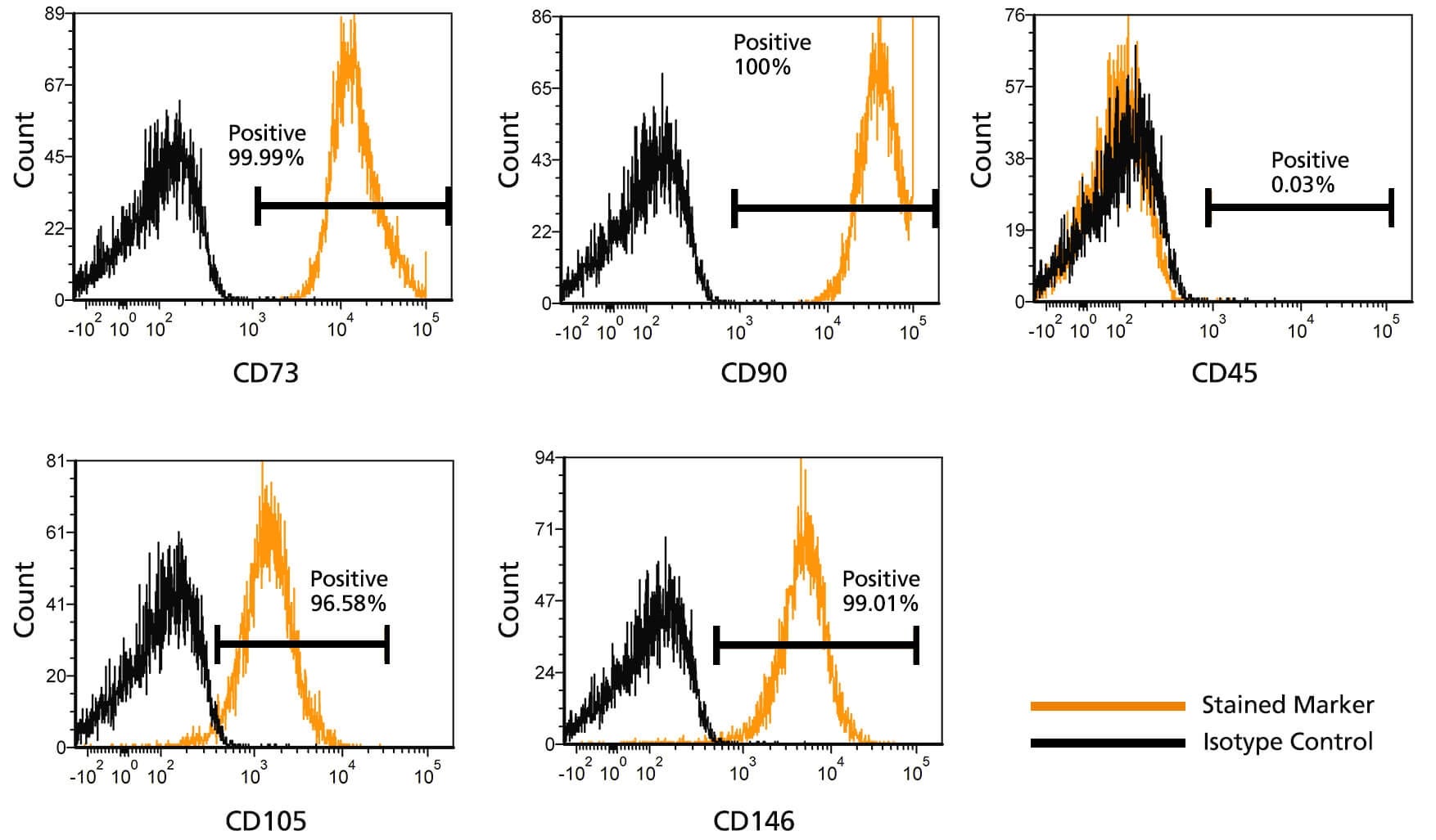
Figure 4. Flow Cytometric Analysis of MSCs Cultured in MesenCult™-ACF Plus Medium.
BM-derived MSCs were cultured and expanded in MesenCult™-ACF Plus Medium. At passage 8 MSCs were stained for mesenchymal surface markers (CD73, CD90, CD105,), pericyte marker (CD146) and hematopoietic marker (CD45). MSCs expressed high levels of CD73, CD90, CD105 and CD146 and lacked expression of CD45.

产品说明书及文档

请在《产品说明书》中查找相关支持信息和使用说明,或浏览下方更多实验方案。

Document Type
Product Name
Catalog #
Lot #
Language
Catalog #
05448
Lot #
All
Language
English
Document Type
Safety Data Sheet 1
Catalog #
05448
Lot #
All
Language
English
Document Type
Safety Data Sheet 2
Catalog #
05448
Lot #
All
Language
English
Document Type
Safety Data Sheet 3
Catalog #
05448
Lot #
All
Language
English

应用领域

本产品专为以下研究领域设计,适用于工作流程中的高亮阶段。探索这些工作流程,了解更多我们为各研究领域提供的其他配套产品。

相关材料与文献

技术资料 (13)

文献 (11)

Characterization and Therapeutic Application of Mesenchymal Stem Cells with Neuromesodermal Origin from Human Pluripotent Stem Cells. H. Wang et al. Theranostics 2019

Abstract

Rationale: Mesenchymal stem cells (MSC) hold great promise in the treatment of various diseases including autoimmune diseases,inflammatory diseases,etc.,due to their pleiotropic properties. However,largely incongruent data were obtained from different MSC-based clinical trials,which may be partially due to functional heterogeneity among MSC. Here,we attempt to derive homogeneous mesenchymal stem cells with neuromesodermal origin from human pluripotent stem cells (hPSC) and evaluate their functional properties. Methods: Growth factors and/or small molecules were used for the differentiation of human pluripotent stem cells (hPSC) into neuromesodermal progenitors (NMP),which were then cultured in animal component-free and serum-free induction medium for the derivation and long-term expansion of MSC. The resulted NMP-MSC were detailed characterized by analyzing their surface marker expression,proliferation,migration,multipotency,immunomodulatory activity and global gene expression profile. Moreover,the in vivo therapeutic potential of NMP-MSC was detected in a mouse model of contact hypersensitivity (CHS). Results: We demonstrate that NMP-MSC express posterior HOX genes and exhibit characteristics similar to those of bone marrow MSC (BMSC),and NMP-MSC derived from different hPSC lines show high level of similarity in global gene expression profiles. More importantly,NMP-MSC display much stronger immunomodulatory activity than BMSC in vitro and in vivo,as revealed by decreased inflammatory cell infiltration and diminished production of pro-inflammatory cytokines in inflamed tissue of CHS models. Conclusion: Our results identify NMP as a new source of MSC and suggest that functional and homogeneous NMP-MSC could serve as a candidate for MSC-based therapies.
Glycyrrhetinic Acid Maintains Intestinal Homeostasis via HuR. G. Chen et al. Frontiers in pharmacology 2019

Abstract

Glycyrrhetinic acid (GA) is one of the main components of the traditional Chinese medicine of licorice,which can coordinate and promote the effects of other medicines in the traditional prescription. We found that GA could promote the proliferation,decrease the apoptotic rate,and attenuate DFMO-elicited growth arrest and delay in restitution after wounding in IEC-6 cells via HuR. GA failed to promote proliferation and to suppress apoptosis after silencing HuR by siRNA in IEC-6 cells. Furthermore,with the model of small intestinal organoids developed from intestinal crypt stem cells,we found that GA could increase HuR and its downstream ki67 levels to promote intestinal organoid development. In the in vivo assay,GA was shown to maintain the integrity of the intestinal epithelium under the circumstance of 48 h-fasting in rats via raising HuR and its downstream genes such as EGF,EGFR,and MEK. These results suggested that via HuR modulation,GA could promote intestinal epithelium homeostasis,and therefore contribute to the absorption of constituents from other medicines co-existing in the traditional prescription with licorice in the small intestine. Our results provide a new perspective for understanding the effect of licorice on enhancing the therapeutic effect of traditional prescriptions according to the traditional Chinese medicine theory.
Gestational Tissue-Derived Human Mesenchymal Stem Cells Use Distinct Combinations of Bioactive Molecules to Suppress the Proliferation of Human Hepatoblastoma and Colorectal Cancer Cells. N. Paiboon et al. Stem cells international 2019

Abstract

Background Cancer has been considered a serious global health problem and a leading cause of morbidity and mortality worldwide. Despite recent advances in cancer therapy,treatments of advance stage cancers are mostly ineffective resulting in poor survival of patients. Recent evidences suggest that multipotent human mesenchymal stem cells (hMSCs) play important roles in growth and metastasis of several cancers by enhancing their engraftment and inducing tumor neovascularization. However,the effect of hMSCs on cancer cells is still controversial because there are also evidences demonstrating that hMSCs inhibited growth and metastasis of some cancers. Methods In this study,we investigated the effects of bioactive molecules released from bone marrow and gestational tissue-derived hMSCs on the proliferation of various human cancer cells,including C3A,HT29,A549,Saos-2,and U251. We also characterized the hMSC-derived factors that inhibit cancer cell proliferation by protein fractionation and mass spectrometry analysis. Results We herein make a direct comparison and show that the effects of hMSCs on cancer cell proliferation and migration depend on both hMSC sources and cancer cell types and cancer-derived bioactive molecules did not affect the cancer suppressive capacity of hMSCs. Moreover,hMSCs use distinct combination of bioactive molecules to suppress the proliferation of human hepatoblastoma and colorectal cancer cells. Using protein fractionation and mass spectrometry analysis,we have identified several novel hMSC-derived factors that might be able to suppress cancer cell proliferation. Conclusion We believe that the procedure developed in this study could be used to discover other therapeutically useful molecules released by various hMSC sources for a future in vivo study.

更多信息

更多信息
物种
配方 不含动物成分, 无血清
质量保证:

产品仅供研究使用,不用于针对人或动物的诊断或治疗。 欲获悉更多关于STEMCELL的质控信息,请访问 STEMCELL.CN/COMPLIANCE.
Copyright © 2026 by STEMCELL Technologies. All rights reserved.

在线联系