
 EasySep™小鼠TIL(CD45)正选试剂盒


产品号 #05240_C
用于分化和扩增间充质祖细胞的成分明确的培养试剂盒
若您需要咨询产品或有任何技术问题,请通过官方电话 400 885 9050 或邮箱 info.cn@stemcell.com 与我们联系。
用于分化和扩增间充质祖细胞的成分明确的培养试剂盒
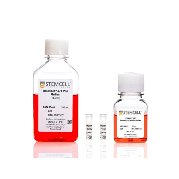
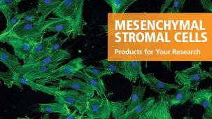
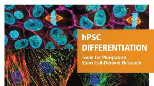
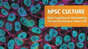
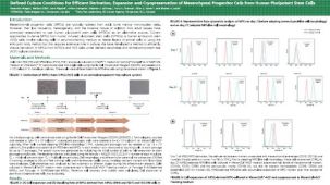
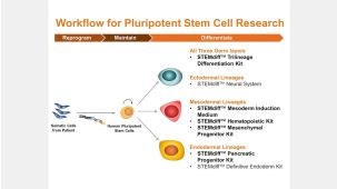
使用STEMdiff™间充质祖细胞试剂盒,可高效且可重复地从人多能干细胞(hPSCs)中诱导生成间充质祖细胞(MPCs)。这款完全无动物成分(ACF)的试剂盒提供从人胚胎干细胞(ESCs)或诱导多能干细胞(iPSCs)诱导和扩增MPCs所需的所有试剂。
使用该试剂盒生成的细胞表达标志性的间充质标志物(CD73、CD90、CD105),当与MesenCult™分化试剂盒配合使用时,表现出强效扩增以及向脂肪生成、成骨和软骨生成谱系的分化能力。与原代间充质基质细胞(MSCs)相比,使用该工作流程衍生的MPCs表现出降低的异质性,并为再生医学研究、疾病建模和药物发现提供了一致且可重复的平台。
有关MSC的进一步分化和培养,请查看我们的MesenCult™ MSC细胞培养产品组合,其中包含人iPSC衍生的间充质祖细胞,以便您使用经过验证的纯MSC细胞培养物开始您的实验。
CollPlant是细胞贴附基质中重组人胶原蛋白(rhCollagen)成分的制造商。
本产品仅供研究使用。如需将本产品用于任何临床或商业用途,请联系STEMCELL。
分类
专用培养基
细胞类型
间充质细胞,PSC衍生
种属
人
应用
细胞培养,分化
品牌
STEMdiff
研究领域
药物发现和毒性检测,干细胞生物学
制剂类别
不含动物成分,无血清
请在《产品说明书》中查找相关支持信息和使用说明,或浏览下方更多实验方案。
本产品专为以下研究领域设计,适用于工作流程中的高亮阶段。探索这些工作流程,了解更多我们为各研究领域提供的其他配套产品。
| 物种 | 人 |
|---|---|
| 配方 | 不含动物成分, 无血清 |
人MSCs向脂肪细胞分化的培养基
用于MSCs向软骨细胞分化的无动物成分培养基
在线联系


 沪公网安备31010102008431号