
 EasySep™小鼠TIL(CD45)正选试剂盒


产品号 #(选择产品)
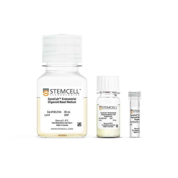
用于扩增和分化人子宫内膜类器官的无血清、无酚红、无性类固醇激素培养基。
若您需要咨询产品或有任何技术问题,请通过官方电话 400 885 9050 或邮箱 info.cn@stemcell.com 与我们联系。
用于扩增和分化人子宫内膜类器官的无血清、无酚红、无性类固醇激素培养基。
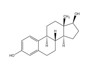
使用GyneCult™子宫内膜类器官培养基(EOM)生成对激素具有响应性的子宫内膜类器官。该培养基不含血清、酚红和性类固醇激素,专为支持女性生殖生物学研究而设计。在培养过程中,可向GyneCult™ EOM中添加不同浓度的雌激素和/或孕激素,以灵活模拟月经周期的不同阶段。生成的类器官对激素有反应,并包含体内主要上皮细胞类型和标志物,例如孕激素相关子宫内膜蛋白(PAEP)、乙酰化微管蛋白、PAX8和SOX9。
该优化的培养基支持从多种样本来源(包括上皮细胞含量较低的样本)稳定地构建类器官,并且与来自增殖期和分泌期子宫内膜、子宫内膜异位症病灶和经期样本的原代细胞兼容。该培养基成分明确、可生成生理相关的细胞类型、样本来源广泛,可在受控的激素调节下灵活地模拟健康和病变的子宫内膜状态,有助于填补目前子宫内膜和女性生殖健康研究方面的不足。
细胞类型
上皮细胞
应用
细胞培养、分化、类器官培养
品牌
GyneCult
研究领域
癌症、疾病建模、药物发现与毒性检测、上皮细胞生物学、类器官
配方
无血清
请在《产品说明书》中查找相关支持信息和使用说明,或浏览下方更多实验方案。
本产品专为以下研究领域设计,适用于工作流程中的高亮阶段。探索这些工作流程,了解更多我们为各研究领域提供的其他配套产品。
| 配方 | 无血清 |
|---|
用于在气液界面培养的人呼吸道上皮细胞的无血清和无BPE培养基
用于 CFU 检测人乳腺上皮细胞的培养
用于培养人、小鼠和大鼠上皮干细胞

<p>用于扩增和分化人输卵管类器官的无血清、支持性激素调节的试剂盒</p>
法律声明:
本产品仅供研究使用,除非另有说明,不得用于人类或动物的诊断或治疗用途。如需了解STEMCELL质量的更多信息,请参阅 WWW.STEMCELL.COM/COMPLIANCE。
安全声明:
CA WARNING: This product can expose you to Ethyl Alcohol which is known to the State of California to cause cancer and birth defects or other reproductive harm. For more information go to www.P65Warnings.ca.gov
在线联系


 沪公网安备31010102008431号