
 EasySep™小鼠TIL(CD45)正选试剂盒


产品号 #19669_C
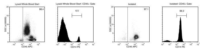
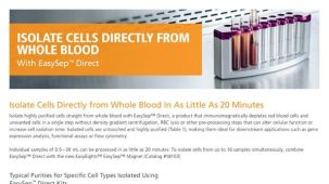
直接从全血中免疫磁珠负选人单核细胞
若您需要咨询产品或有任何技术问题,请通过官方电话 400 885 9050 或邮箱 info.cn@stemcell.com 与我们联系。
直接从全血中免疫磁珠负选人单核细胞
使用EasySep™ Direct人单核细胞分选试剂盒,可轻松高效地从人全血样本中通过免疫磁珠负选获得高纯度人CD14+单核细胞。EasySep™技术结合单克隆抗体的特异性和免磁柱系统的简便性,已在发表的研究中广泛应用超过20年。
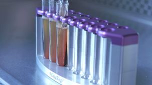
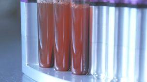
在该EasySep™负选流程中,非目的细胞会被抗体复合物和EasySep™ Direct RapidSpheres™磁珠标记。以下非目的 细胞会被特异性去除:粒细胞、T细胞、B细胞、NK细胞、树突状细胞、血小板和红系细胞。通过EasySep™磁极将被磁珠标记的细胞与未被标记的目的细胞分离,目的细胞可被轻松收集到新试管中,分选后的细胞可立即用于下游应用,例如流式细胞术、培养或DNA/RNA 提取。
了解更多EasySep™免疫磁珠 技术的工作原理,或者如何通过RoboSep™实现全自动化免疫磁珠细胞分选。探索更多为您的实验流程优化的产品,包括细胞鉴定 、冷冻保存等相关试剂 。
磁极兼容性
• EasySep™磁极(产品号 #18000)
• “The Big Easy” EasySep™磁极(产品号 #18001)
• Easy 50 EasySep™磁极(产品号 #18002)
• EasyEights™ EasySep™磁极(产品号 #18103)
• RoboSep™-S(产品号 #21000)
分类
细胞分选试剂盒
细胞类型
单核细胞,髓系细胞
种属
人
样本来源
全血
分选方法
负选
应用
细胞分选
品牌
EasySep,RoboSep
研究领域
药物发现和毒性检测,免疫学
请在《产品说明书》中查找相关支持信息和使用说明,或浏览下方更多实验方案。
本产品专为以下研究领域设计,适用于工作流程中的高亮阶段。探索这些工作流程,了解更多我们为各研究领域提供的其他配套产品。
| 物种 | 人 |
|---|---|
| Magnet Compatibility | • EasySep™ Magnet (Catalog #18000) • “The Big Easy” EasySep™ Magnet (Catalog #18001) • Easy 50 EasySep™ Magnet (Catalog #18002) • EasyEights™ EasySep™ Magnet (Catalog #18103) • RoboSep™-S (Catalog #21000) |
| 样本来源 | 全血 |
| Selection Method | Negative |
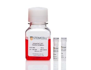
人单核细胞向树突状细胞分化的完整试剂盒
通过免疫磁珠负选分离无磁珠标记的人CD14+CD16-单核细胞
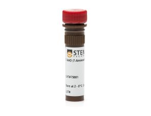
细胞活性染料(DNA标记染料)

细胞活力染料(DNA标记染料)
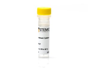
粒细胞-巨噬细胞集落刺激因子
小鼠Monoclonal IgG1抗体,抗人、黑猩猩CD45
抗人、恒河猴、食蟹猴CD14的小鼠Monoclonal IgG2a抗体
在线联系


 沪公网安备31010102008431号