若您需要咨询产品或有任何技术问题,请通过官方电话 400 885 9050 或邮箱 info.cn@stemcell.com 与我们联系。

EasySep™ 死细胞去除 (Annexin V) 试剂盒

免疫磁珠去除凋亡细胞(Annexin V)

只有 %1
¥5,142.00

产品号 #(选择产品)

产品号 #17899_C

免疫磁珠去除凋亡细胞(Annexin V)

产品优势

  • 快捷、操作简单
  • 无需分离柱
  • 兼容EasySep™、“The Big Easy”和EasyEights™磁极

产品组分包括

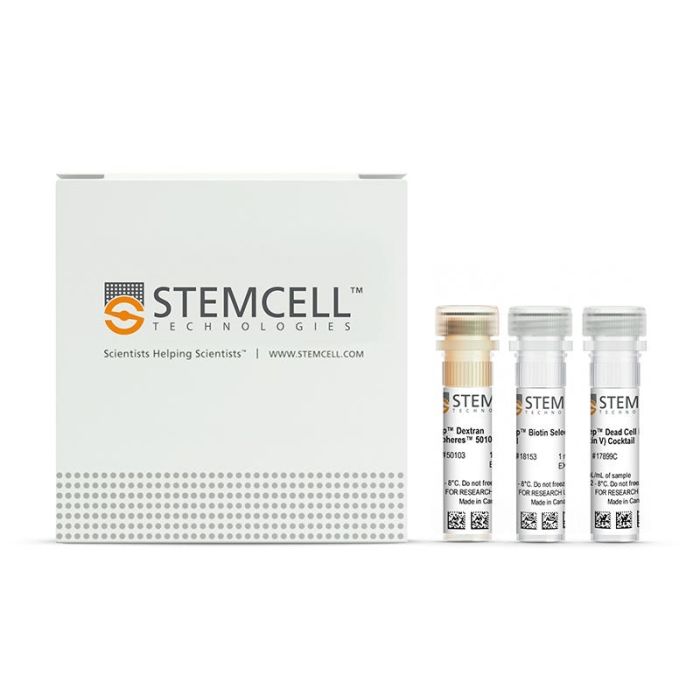
  • EasySep™死细胞去除(Annexin V)试剂盒(产品号 #17899)
    • EasySep™死细胞去除(Annexin V)抗体复合物, 0.5 mL
    • EasySep™生物素分选抗体复合物,1mL
    • EasySep™Dextran RapidSpheres™50103磁珠,1 mL
专为您的实验方案打造的产品
要查看实验方案所需的所有配套产品,请参阅《实验方案与技术文档》

总览

使用 EasySep™ 死细胞去除 (Annexin V) 试剂盒,通过免疫磁性负选,可高效去除细胞培养或组织制备样本中的凋亡细胞 (Annexin V+)。EasySep™结合了单克隆抗体的特异性和无柱磁性系统的简便性,迄今已广泛应用于发表的研究中超过20年。

EasySep™ 操作流程简单,使用识别 Annexin V的抗体复合物和磁珠标记细胞,再通过 EasySep™ 磁极进行无柱分选,只需倾倒或吸取未标记细胞,即可将标记细胞与未标记细胞分离,Annexin V+ 细胞则保留在管中。经过磁性细胞分离后,即可获得所需的细胞用于下游应用。在细胞凋亡过程中,Annexin V 会与细胞膜外层磷脂酰丝氨酸结合。

了解更多关于免疫磁性 EasySep™ 技术的工作原理。探索更多优化您实验流程的产品,包括培养基、补充剂、抗体等。

磁极兼容性
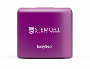
• EasySep™磁极(产品号 #18000),或
• “The Big Easy” EasySep™磁极(产品号 #18001),或
• EasyEights™ EasySep™磁极(产品号 #18103)
 
分类
细胞分选试剂盒
 
细胞类型
淋巴细胞
 
种属
人,小鼠,非人灵长类,其他组织,大鼠
 
样本来源
脐带血,白细胞单采术样本,肺,淋巴结,其他组织,脾脏
 
分选方法
去除
 
品牌
EasySep
 
研究领域
免疫
 

实验数据

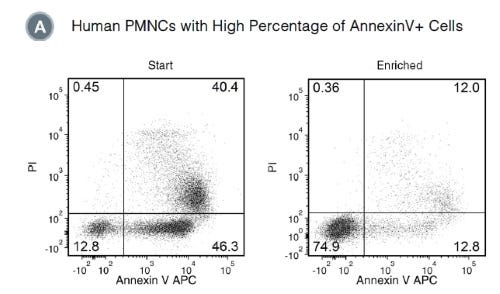
Figure 1. Typical Profile for Dead Cell Removal from Human PMNCs Using EasySep™ Dead Cell Removal (Annexin V) Kit

Starting with human polymorphonuclear cells (PMNCs) cultured overnight, the live cell content (AnnexinV-/PI-) of the enriched fraction is typically 69.7± 12.5% (mean ± SD), using the purple EasySep™ Magnet. In the above example, the percentages of live cells in the start and final enriched fractions are 12.8% and 74.9%, respectively.

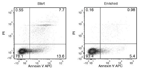
Figure 2. Typical Profile for Dead Cell Removal from Mouse Splenocytes Using EasySep™ Dead Cell Removal (Annexin V) Kit

Starting with 24- to 48-hour-old mouse splenocytes, the live cell content of the enriched fraction is typically 79.8 ± 11.4% (mean ± SD), using the purple EasySep™ Magnet. In the above example, the percentages of live cells in the start and final enriched fractions are 78.1% and 93.4%, respectively.

产品说明书及文档

请在《产品说明书》中查找相关支持信息和使用说明,或浏览下方更多实验方案。

Document Type
Product Name
Catalog #
Lot #
Language
Document Type
产品说明书
Catalog #
17899
Lot #
All
Language
中文
Catalog #
17899
Lot #
All
Language
English
Document Type
Safety Data Sheet 1
Catalog #
17899
Lot #
All
Language
English
Document Type
Safety Data Sheet 2
Catalog #
17899
Lot #
All
Language
English
Document Type
Safety Data Sheet 3
Catalog #
17899
Lot #
All
Language
English

相关材料与文献

技术资料 (8)

文献 (21)

Exosome mimicry by a HAVCR1-NPC1 pathway of endosomal fusion mediates hepatitis A virus infection. M. I. Costafreda et al. Nature microbiology 2020 jun

Abstract

Cell-to-cell communication by exosomes controls normal and pathogenic processes1,2. Viruses can spread in exosomes and thereby avoid immune recognition3. While biogenesis,binding and uptake of exosomes are well characterized4,5,delivery of exosome cargo into the cytoplasm is poorly understood3. We report that the phosphatidylserine receptor HAVCR1 (refs. 6,7) and the cholesterol transporter NPC1 (ref. 8) participate in cargo delivery from exosomes of hepatitis A virus (HAV)-infected cells (exo-HAV) by clathrin-mediated endocytosis. Using CRISPR-Cas9 knockout technology,we show that these two lipid receptors,which interact in the late endosome9,are necessary for the membrane fusion and delivery of RNA from exo-HAV into the cytoplasm. The HAVCR1-NPC1 pathway,which Ebola virus exploits to infect cells9,mediates HAV infection by exo-HAV,which indicates that viral infection via this exosome mimicry mechanism does not require an envelope glycoprotein. The capsid-free viral RNA in the exosome lumen,but not the endosomal uncoating of HAV particles contained in the exosomes,is mainly responsible for exo-HAV infectivity as assessed by methylene blue inactivation of non-encapsidated RNA. In contrast to exo-HAV,infectivity of HAV particles is pH-independent and requires HAVCR1 or another as yet unidentified receptor(s) but not NPC1. Our findings show that envelope-glycoprotein-independent fusion mechanisms are shared by exosomes and viruses,and call for a reassessment of the role of envelope glycoproteins in infection.
A Pygopus 2-Histone Interaction Is Critical for Cancer Cell Dedifferentiation and Progression in Malignant Breast Cancer. M. Saxena et al. Cancer research 2020 sep

Abstract

Pygopus 2 (Pygo2) is a coactivator of Wnt/$\beta$-catenin signaling that can bind bi- or trimethylated lysine 4 of histone-3 (H3K4me2/3) and participate in chromatin reading and writing. It remains unknown whether the Pygo2-H3K4me2/3 association has a functional relevance in breast cancer progression in vivo. To investigate the functional relevance of histone-binding activity of Pygo2 in malignant progression of breast cancer,we generated a knock-in mouse model where binding of Pygo2 to H3K4me2/3 was rendered ineffective. Loss of Pygo2-histone interaction resulted in smaller,differentiated,and less metastatic tumors,due,in part,to decreased canonical Wnt/$\beta$-catenin signaling. RNA- and ATAC-sequencing analyses of tumor-derived cell lines revealed downregulation of TGF$\beta$ signaling and upregulation of differentiation pathways such as PDGFR signaling. Increased differentiation correlated with a luminal cell fate that could be reversed by inhibition of PDGFR activity. Mechanistically,the Pygo2-histone interaction potentiated Wnt/$\beta$-catenin signaling,in part,by repressing the expression of Wnt signaling antagonists. Furthermore,Pygo2 and $\beta$-catenin regulated the expression of miR-29 family members,which,in turn,repressed PDGFR expression to promote dedifferentiation of wild-type Pygo2 mammary epithelial tumor cells. Collectively,these results demonstrate that the histone binding function of Pygo2 is important for driving dedifferentiation and malignancy of breast tumors,and loss of this binding activates various differentiation pathways that attenuate primary tumor growth and metastasis formation. Interfering with the Pygo2-H3K4me2/3 interaction may therefore serve as an attractive therapeutic target for metastatic breast cancer. SIGNIFICANCE: Pygo2 represents a potential therapeutic target in metastatic breast cancer,as its histone-binding capability promotes $\beta$-catenin-mediated Wnt signaling and transcriptional control in breast cancer cell dedifferentiation,EMT,and metastasis.
Cancer cell-intrinsic resistance to BiTE therapy is mediated by loss of CD58 costimulation and modulation of the extrinsic apoptotic pathway. Y. Shen et al. Journal for immunotherapy of cancer 2022 mar

Abstract

BACKGROUND Bispecific T-cell engager (BiTE) molecules induce redirected lysis of cancer cells by T cells and are an emerging modality for solid tumor immunotherapy. While signs of clinical activity have been demonstrated,efficacy of T-cell engagers (TCEs) in solid tumors settings,molecular determinants of response,and underlying mechanisms of resistance to BiTE therapy require more investigation. METHODS To uncover cancer cell-intrinsic genetic modifiers of TCE-mediated cytotoxicity,we performed genome-wide CRISPR (Clustered Regularly Interspaced Short Palindromic Repeats) loss-of-function and CRISPRa (CRISPR activation) gain-of-function screens using TCEs against two distinct tumor-associated antigens (TAAs). By using in vitro T-cell cytotoxicity assays and in vivo efficacy studies,we validated the roles of two common pathways identified in our screen,T-cell costimulation pathway and apoptosis pathway,as key modifiers of BiTE activity. RESULTS Our genetic screens uncovered TAAs-independent cancer cell-intrinsic genes with functions in autophagy,T-cell costimulation,the apoptosis pathway,chromatin remodeling,and cytokine signaling that altered responsiveness to BiTE-mediated killing. Notably,loss of CD58 (the ligand of the CD2 T-cell costimulatory receptor),a gene frequently altered in cancer,led to decreased TCE-mediated cytotoxicity,T-cell activation and antitumor efficacy in vitro and in vivo. Moreover,the effects of CD58 loss were synergistically compounded by concurrent loss of CD80/CD86 (ligands for the CD28 T-cell costimulatory receptor),whereas joint CD2 and CD28 costimulation additively enhanced TCE-mediated killing,indicating non-redundant costimulatory mechanisms between the two pathways. Additionally,loss of CFLAR (Caspase-8 and FADD Like Apoptosis Regulator),BCL2L1,and BID (BH3 Interacting Domain Death Agonist) induced profound changes in sensitivity to TCEs,indicating that key regulators of apoptosis,which are frequently altered in cancer,impact tumor responsiveness to BiTE therapy. CONCLUSIONS This study demonstrates that genetic alterations central to carcinogenesis and commonly detected in cancer samples lead to significant modulation of BiTE antitumor activity in vitro and in vivo,findings with relevance for a better understanding of patient responses to BiTE therapy and novel combinations that enhance TCE efficacy.

更多信息

更多信息
物种 人, 其它物种, 大鼠, 小鼠, 非人灵长类
Magnet Compatibility • EasySep™ Magnet (Catalog #18000), or • “The Big Easy” EasySep™ Magnet (Catalog #18001), or • EasyEights™ EasySep™ Magnet (Catalog #18103)
样本来源 其它细胞系, 淋巴结, 白细胞单采术样本, 肺, 脐带血, 脾脏
Selection Method Depletion
质量保证:

产品仅供研究使用,不用于针对人或动物的诊断或治疗。 欲获悉更多关于STEMCELL的质控信息,请访问 STEMCELL.CN/COMPLIANCE.
Copyright © 2026 by STEMCELL Technologies. All rights reserved.

在线联系