若您需要咨询产品或有任何技术问题,请通过官方电话 400 885 9050 或邮箱 info.cn@stemcell.com 与我们联系。

抗小鼠SSEA-1(CD15)抗体,clone MC-480

抗人、小鼠、大鼠SSEA-1(CD15)的小鼠Monoclonal IgM抗体
只有 %1
¥2,190.00

产品号 #(选择产品)

产品号 #60060_C

抗人、小鼠、大鼠SSEA-1(CD15)的小鼠单克隆IgM抗体

总览

MC-480抗体与末端碳水化合物表位,阶段特异性胚胎抗原-1 (SSEA-1)反应,SSEA-1表达于小鼠早期胚胎,小鼠胚胎癌(EC),胚胎干细胞(ES)和小鼠及人胚胎生殖(EG)细胞表面的一种大分子量(> 200 kDa)糖蛋白上。SSEA-1在未分化的人EC、ES或诱导多能干细胞(iPS)或恒河猴ES细胞系上不表达。其在小鼠胚胎干细胞中的表达在分化过程中降低,而在人ES细胞中,其表达在分化过程中会上调。SSEA-1也存在于成人粒细胞和单核细胞中,标记为CD15, MC-480抗体识别这些细胞类型上的CD15标记。据报道,SSEA-1在细胞粘附和迁移以及细胞分化调控中发挥作用。

该抗体Clone已通过EasySep™试剂盒分离的细胞纯度评估验证,包括EasySep™HLA全血CD15正选试剂盒(产品号#18681HLA;可观察到部分阻断),并用于标记在TeSR™-E8™(产品号#05940),mTeSR™1(产品号#85850)和TeSR™2(产品号#05860)中生长的人 ES和iPS细胞。

分类
一抗
 
靶抗原
SSEA-1 (CD15)
 
别名
3-FAL,CD15,Lewis X,SSEA1,阶段特异性胚胎抗原1,X-半抗原
 
反应种属
人,小鼠,大鼠
 
偶联
Alexa Fluor 488,生物素,FITC,PE,未偶联
 
宿主物种
小鼠
 
细胞类型
多能干细胞
 
种属
人,小鼠,大鼠
 
应用
细胞分选,流式细胞术,免疫细胞化学,免疫荧光,免疫组化,免疫沉淀,Western印迹
 
研究领域
干细胞生物学
 
Clone
MC-480
 
Gene ID
14345
 
Isotype
IgM, κ
 

实验数据

Data for Alexa Fluor® 488-Conjugated

Figure 1. Data for Alexa Fluor® 488-Conjugated

(A) Flow cytometry analysis of human whole blood nucleated cells labeled with Anti-Human SSEA-1 (CD15) Antibody, Clone MC-480, Alexa Fluor® 488 (filled histogram) or Mouse IgM, kappa Isotype Control Antibody, Clone MM-30, Alexa Fluor® 488 (Catalog #60069AD) (solid line histogram). SSEA-1 is highly expressed on granulocytes.
(B) Flow cytometry analysis of human HT1080 fibrosarcoma cells labeled with Anti-Human SSEA-1 (CD15) Antibody, Clone MC-480, Alexa Fluor® 488 (filled histogram). Labeling of human HT1080 fibrosarcoma (solid line histogram) or ES cells (negative control; dashed line histogram) with a mouse IgM, kappa isotype control antibody (Anti-Human TRA-1-60 Antibody, Clone TRA-1-60R, Alexa Fluor® 488; Catalog #60064AD) is shown. SSEA-1 is not expressed on undifferentiated human ES cells.
(C) Human ES cells were cultured in mTeSR™1 on Corning® Matrigel®-coated glass slides, then fixed and labeled with Anti-Human SSEA-1 (CD15) Antibody, Clone MC-480, Alexa Fluor® 488. Inset shows cells labeled with Mouse IgM, kappa Isotype Control Antibody, Clone MM-30, Alexa Fluor® 488.

Data for Biotin-Conjugated

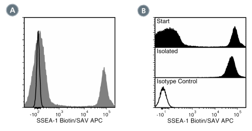
Figure 2. Data for Biotin-Conjugated

(A) Flow cytometry analysis of human buffy coat nucleated cells labeled with Anti-Human SSEA-1 Antibody, Clone MC-480, Biotin, followed by streptavidin (SAV) APC (filled histogram), or a mouse IgM, kappa biotin isotype control antibody, followed by SAV APC (solid line histogram). (B) Flow cytometry analysis of human buffy coat nucleated cells processed with the EasySep™ HLA Whole Blood CD15 Positive Selection Kit and labeled with Anti-Human SSEA-1 Antibody, Clone MC-480, Biotin, followed by SAV APC. Histograms show labeling of buffy coat nucleated cells (Start) and isolated cells (Isolated). Labeling with a mouse IgM, kappa biotin isotype control antibody, followed by SAV APC is shown (solid line histogram).

Data for FITC-Conjugated

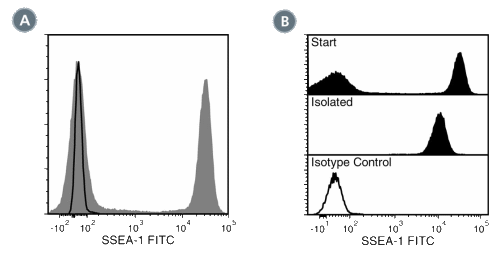
Figure 3. Data for FITC-Conjugated

(A) Flow cytometry analysis of human buffy coat nucleated cells labeled with Anti-Human SSEA-1 Antibody, Clone MC-480, FITC (filled histogram) or a mouse IgM, kappa FITC Isotype control antibody (solid line histogram). SSEA-1 (CD15) is highly expressed on granulocytes. (B) Flow cytometry analysis of human buffy coat nucleated cells processed with the EasySep™ HLA CD15 WB Positive Selection Kit and labeled with Anti-Human SSEA-1 Antibody, Clone MC-480, FITC. Histograms show labeling of buffy coat nucleated cells (Start) and isolated cells (Isolated). Labeling of start cells with a mouse IgM, kappa FITC Isotype control antibody is shown (solid line histogram).

Data for PE-Conjugated

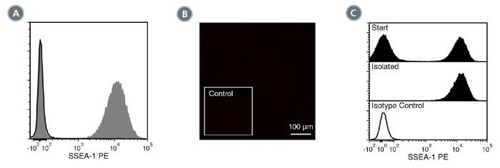
Figure 4. Data for PE-Conjugated

(A) Flow cytometry analysis of human whole blood nucleated cells labeled with Anti-Human SSEA-1 (CD15) Antibody, Clone MC-480, PE (filled histogram) or Mouse IgM, kappa Isotype Control Antibody, Clone MM-30, PE (Catalog #60069PE) (solid line histogram). SSEA-1 is highly expressed on granulocytes.
(B) Human ES cells were cultured in mTeSR™1 on Corning® Matrigel®-coated glass slides, then fixed and labeled with Anti-Human SSEA-1 (CD15) Antibody, Clone MC-480, PE. Inset shows cells labeled with Mouse IgM, kappa Isotype Control Antibody, Clone MM-30, PE. SSEA-1 is not expressed on undifferentiated human ES cells.
(C) Flow cytometry analysis of human buffy coat nucleated cells processed with the EasySep™ HLA CD15 WB Positive Selection Kit and labeled with Anti-Human SSEA-1 (CD15) Antibody, Clone MC-480, PE. Histograms show labeling of buffy coat nucleated cells (Start) and isolated cells (Isolated). Labeling with Mouse IgM, kappa Isotype Control Antibody, Clone MM-30, PE is shown (solid line histogram).

Data for Unconjugated

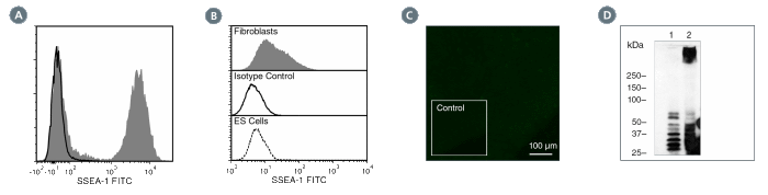
Figure 5. Data for Unconjugated

(A) Flow cytometry analysis of human whole blood nucleated cells labeled with Anti-Human SSEA-1 (CD15) Antibody, Clone MC-480, followed by Goat Anti-Mouse IgG (H+L) Antibody, Polyclonal, FITC (Catalog #60138FI) (filled histogram), or Mouse IgM, kappa Isotype Control Antibody, Clone MM-30 (Catalog #60069), followed by Goat Anti-Mouse IgG (H+L) Antibody, Polyclonal, FITC (solid line histogram). SSEA-1 is highly expressed on granulocytes.
(B) Flow cytometry analysis of human HT1080 fibrosarcoma cells labeled with Anti-Human SSEA-1 (CD15) Antibody, Clone MC-480, followed by goat antimouse IgG, FITC (filled histogram). Labeling of human HT1080 fibrosarcoma cells (solid line histogram) or H1 ES cells (negative control; dashed line histogram) with a mouse IgM, kappa isotype control antibody (Anti-Human TRA-1-60 Antibody, Clone TRA-1-60R; Catalog #60064) is shown.
(C) Human ES cells were cultured in mTeSR™1 on Corning® Matrigel®-coated glass slides, then fixed and labeled with Anti-Human SSEA-1 (CD15) Antibody, Clone MC-480, followed by goat anti-mouse IgG, FITC. Inset shows cells labeled with Mouse IgM, kappa Isotype Control Antibody, Clone MM- 30, followed by goat anti-mouse IgG, FITC. SSEA-1 is not expressed on undifferentiated human ES cells.
(D) Western blot analysis of denatured/reduced cell lysates from human ES cells (negative control; lane 1) or HT1080 fibrosarcoma cells (lane 2) with Anti-Human SSEA-1 (CD15) Antibody, Clone MC-480.

产品说明书及文档

请在《产品说明书》中查找相关支持信息和使用说明,或浏览下方更多实验方案。

Document Type
Product Name
Catalog #
Lot #
Language
Catalog #
60060FI, 60060FI.1
Lot #
All
Language
English
Catalog #
60060AD, 60060AD.1
Lot #
All
Language
English
Catalog #
60060
Lot #
All
Language
English
Catalog #
60060BT
Lot #
All
Language
English
Catalog #
60060PE.1, 60060PE
Lot #
All
Language
English
Document Type
Safety Data Sheet
Catalog #
60060FI, 60060FI.1
Lot #
All
Language
English
Document Type
Safety Data Sheet
Catalog #
60060AD, 60060AD.1
Lot #
All
Language
English
Document Type
Safety Data Sheet
Catalog #
60060
Lot #
All
Language
English
Document Type
Safety Data Sheet
Catalog #
60060BT
Lot #
All
Language
English
Document Type
Safety Data Sheet
Catalog #
60060PE.1, 60060PE
Lot #
All
Language
English

应用领域

本产品专为以下研究领域设计,适用于工作流程中的高亮阶段。探索这些工作流程,了解更多我们为各研究领域提供的其他配套产品。

相关材料与文献

技术资料 (3)

文献 (1)

OCT4 as a target of miR-34a stimulates p63 but inhibits p53 to promote human cell transformation Ng WL et al. Cell death & disease 2014 JAN

Abstract

Human cell transformation is a key step for oncogenic development,which involves multiple pathways; however,the mechanism remains unclear. To test our hypothesis whether cell oncogenic transformation shares some mechanisms with the process of reprogramming non-stem cells to induced pluripotent stem cells (iPSC),we studied the relationship among the key factors for promoting or inhibiting iPSC in radiation-transformed human epithelial cell lines derived from different tissues (lung,breast and colon). We unexpectedly found that p63 and OCT4 were highly expressed (accompanied by low expressed p53 and miR-34a) in all transformed cell lines examined when compared with their non-transformed counterparts. We further elucidated the relationship of these factors: the 3p strand of miR-34a directly targeted OCT4 by binding to the 3′ untranslated region (3′-UTR) of OCT4 and,OCT4,in turn,stimulated p63 but inhibited p53 expression by binding to a specific region of the p63 or p53 promoter. Moreover,we revealed that the effects of OCT4 on promoting cell oncogenic transformation were by affecting p63 and p53. These results support that a positive loop exists in human cells: OCT4 upregulation as a consequence of inhibition of miR-34a,promotes p63 but suppresses p53 expression,which further stimulates OCT4 upregulation by downregulating miR-34a. This functional loop contributes significantly to cell transformation and,most likely,also to the iPSC process.

更多信息

更多信息
物种 人, 大鼠, 小鼠
克隆 MC-480
Gene Id 14345
Alternative Names 3-FAL, CD15, Lewis X, SSEA1, Stage-specific embryonic antigen 1, X-hapten
同种型/isotype IgM, kappa
法律声明:

Alexa Fluor is a registered trademark of Life Technologies Corporation. Antibodies conjugated to Alexa Fluor® are licensed for internal research use only and sale is expressly conditioned on the buyer not using the antibody for manufacturing, performing a service or medical test, or otherwise generating revenue. For use other than research, contact Life Technologies Corporation, 5791 Van Allen Way, Carlsbad, CA 92008 USA or outlicensing@lifetech.com.

质量保证:

产品仅供研究使用,不用于针对人或动物的诊断或治疗。 欲获悉更多关于STEMCELL的质控信息,请访问 STEMCELL.CN/COMPLIANCE.
Copyright © 2026 by STEMCELL Technologies. All rights reserved.

在线联系