
 EasySep™小鼠TIL(CD45)正选试剂盒


产品号 #100-0073_C
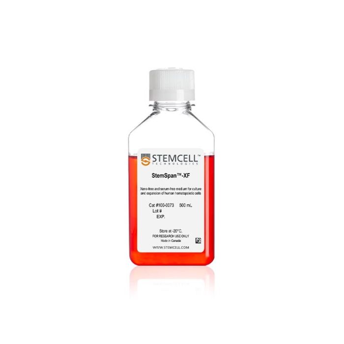
用于培养和扩增人造血细胞的无异源和无血清培养基
若您需要咨询产品或有任何技术问题,请通过官方电话 400 885 9050 或邮箱 info.cn@stemcell.com 与我们联系。
StemSpan™-XF专为体外培养和扩增人造血细胞而开发,可添加适当的生长因子和添加物。这使得用户可以根据实验需求灵活地配制培养基。StemSpan™-XF含有经过测试的人源性和重组人 (rh) 蛋白。
当搭配合适的StemSpan™扩增添加物,StemSpan™-XF可用于扩增从人脐带血、动员外周血或骨髓样本中分离的 CD34+细胞,或扩增和分化谱系定向祖细胞,从而生成红系、髓系或巨核细胞祖细胞群。
包含
本产品仅含有人衍生或重组人蛋白质。
分类
专用培养基
细胞类型
造血干/祖细胞
种属
人
应用
细胞培养,扩增
品牌
StemSpan
研究领域
药物发现和毒性检测,干细胞生物学,移植研究
制剂类别
无血清,无异源
请在《产品说明书》中查找相关支持信息和使用说明,或浏览下方更多实验方案。
本产品专为以下研究领域设计,适用于工作流程中的高亮阶段。探索这些工作流程,了解更多我们为各研究领域提供的其他配套产品。
| 物种 | 人 |
|---|---|
| Contains | This product contains only human-derived or recombinant human proteins. |
| 配方 | 无血清 |
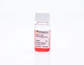
用于扩增人CD34+造血细胞的无血清培养添加物
用于扩增人红系细胞的无血清培养添加物
用于培养和扩增造血细胞的无血清培养基
在线联系


 沪公网安备31010102008431号