
 EasySep™小鼠TIL(CD45)正选试剂盒


产品号 #05110_C
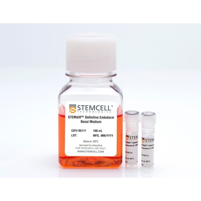
成分明确的无动物成分培养基,用于将人胚胎干细胞(ES)和诱导多能干细胞(iPS)分化为定型内胚层。
若您需要咨询产品或有任何技术问题,请通过官方电话 400 885 9050 或邮箱 info.cn@stemcell.com 与我们联系。
用于将人胚胎干细胞(hESCs)和人诱导多能干细胞(iPSCs)分化为确定性内胚层的定义明确、无动物源成分的培养基
成分明确的无动物成分培养基,用于将人胚胎干细胞(ES)和诱导多能干细胞(iPS)分化为定型内胚层。
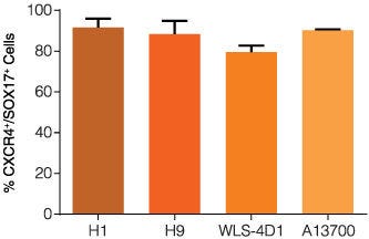
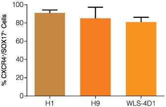
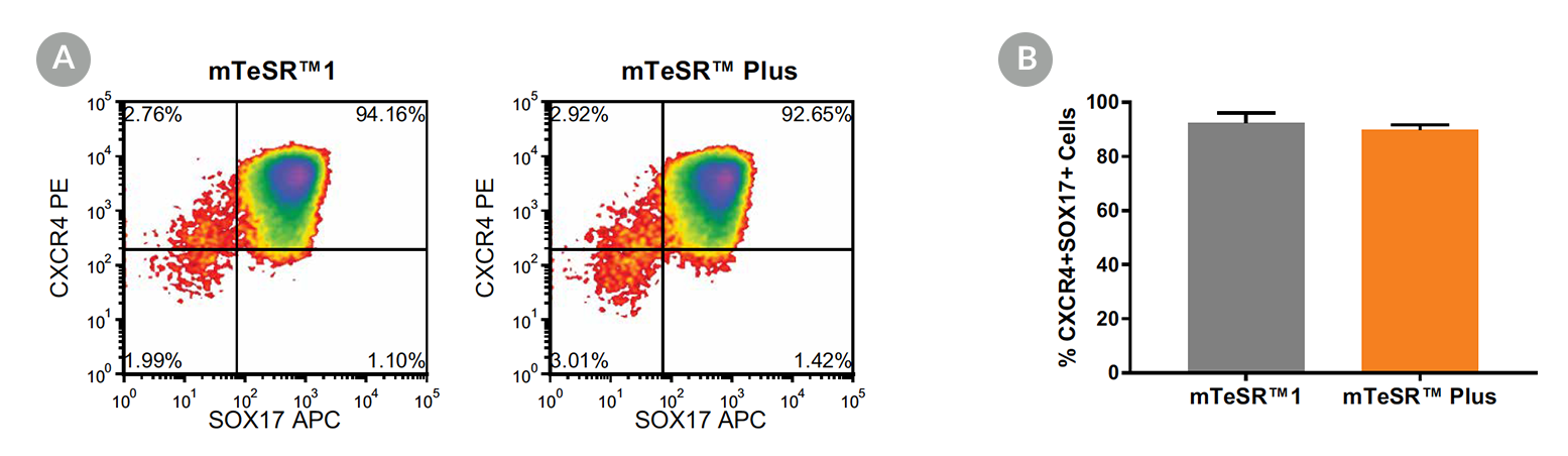
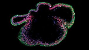
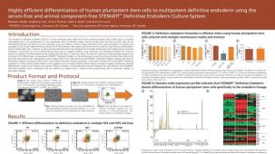
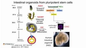
STEMdiff™ Definitive Endoderm Kit 是一套完整的无血清、无动物源成分的培养基与添加物剂盒,可高效支持人胚胎干细胞(hESC)和人诱导多能干细胞(hiPSC)向定向内胚层细胞分化。
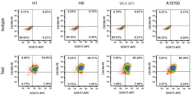
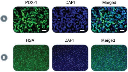
使用 STEMdiff™ Definitive Endoderm Kit 分化的细胞高表达内胚层标志物,包括 CD184(CXCR4)、SOX17、FOXA2 和 c-KIT,同时不表达外胚层、中胚层及多能性标志物。通过该试剂盒获得的定向内胚层细胞具有多向分化潜能,可进一步分化为胰腺、肠道、肺及肝脏谱系细胞,因此是发育生物学研究、疾病建模及药物发现的强大工具。
本试剂盒针对在 mTeSR™1、mTeSR™ Plus 或 TeSR™-AOF 中维持培养的细胞的分化进行了优化。若需了解在 TeSR™-E8™ 中培养的细胞的分化,请参阅 STEMdiff™ Definitive Endoderm Kit (TeSR™-E8™ Optimized)。
分类
专用培养基
细胞类型
气道细胞,内胚层,PSC衍生,肝细胞,肠道细胞,胰腺细胞,多能干细胞
种属
人
应用
细胞培养,分化
品牌
STEMdiff
研究领域
癌症,上皮细胞研究,干细胞生物学
制剂类别
不含动物成分,无血清
请在《产品说明书》中查找相关支持信息和使用说明,或浏览下方更多实验方案。
本产品专为以下研究领域设计,适用于工作流程中的高亮阶段。探索这些工作流程,了解更多我们为各研究领域提供的其他配套产品。
| 物种 | 人 |
|---|---|
| 配方 | 不含动物成分, 无血清 |
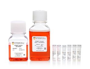
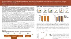
<p>人胚胎干细胞和iPS细胞向胰腺祖细胞分化的无血清培养基</p>
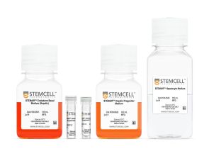
用于将人多能干细胞分化为肝细胞样细胞的无血清分化试剂盒

抗人、食蟹猴、牛CD117(c-Kit)的小鼠Monoclonal IgG1抗体
小鼠Monoclonal IgG2a抗体,抗人、恒河猴、食蟹猴CD184(CXCR4)
质量保证:
产品仅供研究使用,不用于针对人或动物的诊断或治疗。 欲获悉更多关于STEMCELL的质控信息,请访问 STEMCELL.CN/COMPLIANCE.
在线联系

 沪公网安备31010102008431号