
 EasySep™小鼠TIL(CD45)正选试剂盒


技术资料
-
-
-
-
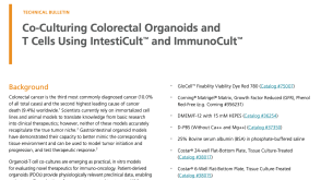 技术公告Co-Culturing Colorectal Organoids and T Cells using IntestiCult™ and ImmunoCult™
细胞类型:
T细胞,肠道细胞
发布日期: 04/01/2024 -
 57:26
线上讲座Consequences of Culture-Acquired Genetic Changes in Human Pluripotent Stem Cells发布日期: 02/20/2024
57:26
线上讲座Consequences of Culture-Acquired Genetic Changes in Human Pluripotent Stem Cells发布日期: 02/20/2024 -
 技术公告Isolating Extracellular Vesicles from Urine with EasySep™ EV Human Positive Selection Kits
细胞类型:
其他细胞系
发布日期: 01/01/2024 -
 技术公告Extracellular Vesicle Generation from Mesenchymal Stromal Cells Using MesenCult™-ACF Plus
细胞类型:
内皮细胞,间充质基质细胞,间充质干祖细胞
发布日期: 12/01/2023


 沪公网安备31010102008431号