
 EasySep™小鼠TIL(CD45)正选试剂盒


技术资料
-
 专家访谈Caroline Lindemans, MD, PhD How Organoids Provide a Model System for Intestinal Regeneration and Repair
研究方向:
上皮细胞生物学,免疫学,干细胞生物学,疾病建模
发布日期: 02/06/2017 -
-
 专家访谈Asier Sáez-Cirión, PhD How Some Patients Can Control HIV Infection
Topics:
- Control of HIV infection
- HIV reservoirs
发布日期: 01/31/2017 -
-
 技术公告Endothelial Protein C Receptor (EPCR): A New Marker for Identification and Positive Selection of Mouse Hematopoietic Stem Cells
细胞类型:
造血干祖细胞
发布日期: 01/09/2017 -
-
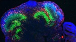
 专家访谈Madeline Lancaster 'Mini-Brains': How an Unexpected Discovery Led to a Groundbreaking Protocol发布日期: 12/22/2016
 -
-
 专家访谈Meritxell Huch, PhD A Drive to Understand Underlying Biologic Principles Made Her an Expert on Hepatic and Pancreatic Organoids
研究方向:
上皮细胞生物学,疾病建模
发布日期: 12/06/2016 -
 专家访谈Tamara Zietek, PhD Studying Intestinal Nutrient Absorption with Organoids
研究方向:
上皮细胞生物学,代谢,疾病建模,癌症研究
发布日期: 12/06/2016

 沪公网安备31010102008431号