
 EasySep™小鼠TIL(CD45)正选试剂盒


技术资料
-
 56:31
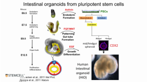
线上讲座Modeling Human Gastrointestinal Development and Disease Using Pluripotent Stem Cells发布日期: 03/06/2017
56:31
线上讲座Modeling Human Gastrointestinal Development and Disease Using Pluripotent Stem Cells发布日期: 03/06/2017 -
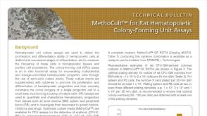 技术公告MethoCult™ for Rat Hematopoietic Colony-Forming Unit (CFU) Assays
细胞类型:
造血干祖细胞
发布日期: 03/01/2017 -
-
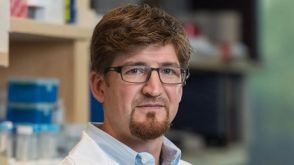 专家访谈Jason Spence Developing New Organoid Systems and the Potential Impact of the Technology
研究方向:
呼吸系统研究
发布日期: 02/21/2017 -
 专家访谈Courteney Lai Life as a Ph.D. Student with a Passion for Myeloid Leukemia
研究方向:
干细胞生物学
发布日期: 02/15/2017 -


 沪公网安备31010102008431号