
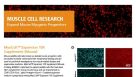
 EasySep™小鼠TIL(CD45)正选试剂盒


技术资料
-
-
-
-
-
-
-
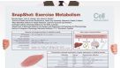 挂图SnapShot: Exercise Metabolism Overview on the skeletal muscle gene expression modulation, energy sources, and inter-organ communications during exercise
 -
-
-

 沪公网安备31010102008431号