
 EasySep™小鼠TIL(CD45)正选试剂盒


技术资料
-
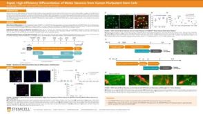 科学海报Rapid, High-Efficiency Differentiation of Motor Neurons from Human Pluripotent Stem Cells
Conference:
FENS 2022
过滤器
筛选结果
- 细胞类型 神经元 删除该内容
研究领域
- 干细胞生物学 1 项目
- 疾病建模 2 项目
- 神经科学 2 项目
- 药物发现和毒理检测 2 项目
Show More
Show Less
产品系列
- BrainPhys 1 项目
- MyoCult 1 项目
- NeuroCult 1 项目
- STEMdiff 1 项目
Show More
Show Less
细胞类型
- B 细胞 236 项目
- CD4+ 46 项目
- CD8+ 29 项目
- CHO细胞 19 项目
- HUVEC细胞(人脐静脉内皮细胞) 1 项目
- NK 细胞 174 项目
- PSC衍生 43 项目
- T 细胞 451 项目
- 上皮细胞 126 项目
- 中胚层 5 项目
- 乳腺细胞 102 项目
- 先天性淋巴细胞 41 项目
- 全血 8 项目
- 其他子集 1 项目
- 其他细胞系 9 项目
- 内皮细胞 12 项目
- 内皮集落形成细胞(ECFCs) 3 项目
- 内胚层 3 项目
- 前列腺细胞 19 项目
- 单个核细胞 92 项目
- 单核细胞 191 项目
- 多能干细胞 1986 项目
- 小胶质细胞 4 项目
- 巨噬细胞 43 项目
- 巨核细胞 10 项目
- 心肌细胞 20 项目
- 成骨细胞 9 项目
- 星形胶质细胞 6 项目
- 杂交瘤细胞 97 项目
- 树突状细胞(DCs) 132 项目
- 气道细胞 4 项目
- 淋巴细胞 83 项目
- 癌细胞和细胞系 1 项目
- 白细胞 17 项目
- 白细胞单采样本 12 项目
- 白血病/淋巴瘤细胞 14 项目
- 监管 1 项目
- 真皮细胞 2 项目
- 神经元 2 项目
- 神经干/祖细胞 472 项目
- 神经细胞 16 项目
- 粒细胞及其亚群 106 项目
- 红系细胞 12 项目
- 红细胞 12 项目
- 肌源干/祖细胞 10 项目
- 肝细胞 34 项目
- 肠道细胞 90 项目
- 肾细胞 4 项目
- 肿瘤细胞 25 项目
- 胰腺细胞 16 项目
- 脂肪细胞 6 项目
- 脑肿瘤干细胞 101 项目
- 血小板 4 项目
- 血浆 3 项目
- 血管生成细胞 4 项目
- 调节性细胞 11 项目
- 软骨细胞 8 项目
- 造血干祖细胞 6 项目
- 造血细胞 4 项目
- 间充质基质细胞 20 项目
- 间充质干祖细胞 1 项目
- 间充质细胞 4 项目
- 骨髓基质细胞 1 项目
- 骨髓间质细胞 1 项目
- 髓系细胞 146 项目
- 肾脏细胞 5 项目
- CD4+T细胞 107 项目
- CD8+T细胞 88 项目
- PSC衍生上皮细胞 30 项目
- PSC衍生中胚层 20 项目
- PSC衍生内皮细胞 12 项目
- PSC衍生内胚层 20 项目
- PSC衍生心肌细胞 21 项目
- PSC衍生神经细胞 116 项目
- PSC衍生肝细胞 11 项目
- PSC衍生造血干细胞 25 项目
- PSC衍生间充质细胞 20 项目
- 其他T细胞亚型 24 项目
- 呼吸道细胞 89 项目
- 多巴胺能神经元 6 项目
- 小鼠胚胎成纤维细胞 1 项目
- 浆细胞 12 项目
- 神经元 191 项目
- 调节性T细胞 64 项目
- 骨髓瘤 5 项目
Show More
Show Less

 沪公网安备31010102008431号