Reuben JM et al. (JUL 2011)
European journal of cancer (Oxford,England : 1990) 47 10 1527--36
Primary breast cancer patients with high risk clinicopathologic features have high percentages of bone marrow epithelial cells with ALDH activity and CD44-CD24lo cancer stem cell phenotype.
BACKGROUND: Cancer stem cells (CSCs) are purported to be epithelial tumour cells expressing CD44(+)CD24(lo) that exhibit aldehyde dehydrogenase activity (Aldefluor(+)). We hypothesised that if CSCs are responsible for tumour dissemination,disseminated cells in the bone marrow (BM) would be positive for putative breast CSC markers. Therefore,we assessed the presence of Aldefluor(+) epithelial (CD326(+)CD45(dim)) cells for the presence of the CD44(+)CD24(lo) phenotype in BM of patients with primary breast cancer (PBC). METHODS: BM aspirates were collected at the time of surgery from 66 patients with PBC. Thirty patients received neoadjuvant chemotherapy (NACT) prior to aspiration. BM was analysed for Aldefluor(+) epithelial cells with or without CD44(+)CD24(lo) expression by flow cytometry. BM aspirates from three healthy donors (HD) were subjected to identical processing and analyses and served as controls. RESULTS: Patients with triple-receptor-negative (TN) tumours had a significantly higher median percentage of CD44(+)CD24(lo) CSC within Aldefluor(+) epithelial cell population than patients with other immunohistochemical subtypes (P=0.018). Patients with TN tumours or with pN2 or higher pathologic nodal status were more likely to have a proportion of CD44(+)CD24(lo) CSC within Aldefluor(+) epithelial cell population above the highest level of HD. Furthermore,patients who received NACT were more likely to have percentages of Aldefluor(+) epithelial cells than the highest level of HD (P=0.004). CONCLUSION: The percentage of CD44(+)CD24(lo) CSC in the BM is higher in PBC patients with high risk tumour features. The selection or enrichment of Aldefluor(+) epithelial cells by NACT may represent an opportunity to target these cells with novel therapies.
View Publication
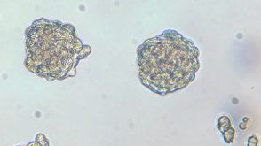
产品号#:
01700
01705
01702
产品名:
ALDEFLUOR™ 试剂盒
ALDEFLUOR™ DEAB试剂, 1.5 mM, 1 mL
ALDEFLUOR™检测缓冲液
Buckley NE et al. (MAR 2011)
Cancer research 71 5 1933--44
The DeltaNp63 proteins are key allies of BRCA1 in the prevention of basal-like breast cancer.
Little is known about the origin of basal-like breast cancers,an aggressive disease that is highly similar to BRCA1-mutant breast cancers. p63 family proteins that are structurally related to the p53 suppressor protein are known to function in stem cell regulation and stratified epithelia development in multiple tissues,and p63 expression may be a marker of basal-like breast cancers. Here we report that ΔNp63 isoforms of p63 are transcriptional targets for positive regulation by BRCA1. Our analyses of breast cancer tissue microarrays and BRCA1-modulated breast cancer cell lines do not support earlier reports that p63 is a marker of basal-like or BRCA1 mutant cancers. Nevertheless,we found that BRCA1 interacts with the specific p63 isoform ΔNp63γ along with transcription factor isoforms AP-2α and AP-2γ. BRCA1 required ΔNp63γ and AP-2γ to localize to an intronic enhancer region within the p63 gene to upregulate transcription of the ΔNp63 isoforms. In mammary stem/progenitor cells,siRNA-mediated knockdown of ΔNp63 expression resulted in genomic instability,increased cell proliferation,loss of DNA damage checkpoint control,and impaired growth control. Together,our findings establish that transcriptional upregulation of ΔNp63 proteins is critical for BRCA1 suppressor function and that defects in BRCA1-ΔNp63 signaling are key events in the pathogenesis of basal-like breast cancer.
View Publication
Reutershan J et al. (MAR 2006)
The Journal of clinical investigation 116 3 695--702
Critical role of endothelial CXCR2 in LPS-induced neutrophil migration into the lung.
In models of acute lung injury,CXC chemokine receptor 2 (CXCR2) mediates migration of polymorphonuclear leukocytes (PMNs) into the lung. Since CXCR2 ligands,including CXCL1 and CXCL2/3,are chemotactic for PMNs,CXCR2 is thought to recruit PMNs by inducing chemotactic migration. In a model of PMN recruitment to the lung,aerosolized bacterial LPS inhalation induced PMN recruitment to the lung in wild-type mice,but not in littermate CXCR2-/- mice. Surprisingly,lethally irradiated wild-type mice reconstituted with CXCR2-/- BM still showed about 50% PMN recruitment into bronchoalveolar lavage fluid and into lung interstitium,but CXCR2-/- mice reconstituted with CXCR2-/- BM showed no PMN recruitment. Conversely,CXCR2-/- mice reconstituted with wild-type BM showed a surprisingly large defect in PMN recruitment,inconsistent with a role of CXCR2 on PMNs alone. Cell culture,immunohistochemistry,flow cytometry,and real-time RT-PCR were used to show expression of CXCR2 on pulmonary endothelial and bronchial epithelial cells. The LPS-induced increase in lung microvascular permeability as measured by Evans blue extravasation required CXCR2 on nonhematopoietic cells. Our data revealed what we believe to be a previously unrecognized role of endothelial and epithelial CXCR2 in LPS-induced PMN recruitment and lung injury.
View Publication
产品号#:
18556
18556RF
产品名:
Xaymardan M et al. (AUG 2009)
Stem cells (Dayton,Ohio) 27 8 1911--20
c-Kit function is necessary for in vitro myogenic differentiation of bone marrow hematopoietic cells.
In recent years,the differentiation of bone marrow cells (BMCs) into myocytes has been extensively investigated,but the findings remain inconclusive. The purpose of this study was to determine the conditions necessary to induce myogenic differentiation in short-term cultures of adult BMCs,and to identify the BMC subpopulation responsible for this phenomenon. We report that high-density cultures of murine hematopoietic BMCs gave rise to spontaneous beating cell clusters in the presence of vascular endothelial and fibroblast growth factors. These clusters originated from c-kit(pos) cells. The formation of the clusters could be completely blocked by adding a c-kit/tyrosine kinase inhibitor,Gleevec (imatinib mesylate; Novartis International,Basel,Switzerland,http://www.novartis.com),to the culture. Cluster formation was also blunted in BMCs from c-kit-deficient (Kit(W)/Kit(W-v)) mice. Clustered cells expressed cardiomyocyte-specific transcription factor genes Gata-4 and Nkx2.5,sarcomeric proteins beta-MHC and MLC-2v,and ANF and connexin-43. Immunostaining revealed alpha-sarcomeric actinin expression in more than 90% of clustered cells. Under electron microscopy,the clustered cells exhibited a sarcomeric myofiber arrangement and z-bands. This study defines the microenvironment required to achieve a reproducible in vitro model of beating,myogenic cell clusters. This model could be used to examine the mechanisms responsible for the postnatal myogenic differentiation of BMCs. Our results identify c-kit(pos) bone marrow hematopoietic cells as the source of the myogenic clusters.
View Publication
产品号#:
18757
18757RF
产品名:
EasySep™小鼠CD117(cKIT)正选试剂盒
RoboSep™ 小鼠CD117(cKIT)正选试剂盒含滤芯吸头
Petersen OW and Polyak K (MAY 2010)
Cold Spring Harbor perspectives in biology 2 5 a003160
Stem cells in the human breast.
The origins of the epithelial cells participating in the development,tissue homeostasis,and cancer of the human breast are poorly understood. However,emerging evidence suggests a role for adult tissue-specific stem cells in these processes. In a hierarchical manner,these generate the two main mammary cell lineages,producing an increasing number of cells with distinct properties. Understanding the biological characteristics of human breast stem cells and their progeny is crucial in attempts to compare the features of normal stem cells and cancer precursor cells and distinguish these from nonprecursor cells and cells from the bulk of a tumor. A historical overview of research on human breast stem cells in primary tissue and in culture reveals the progress that has been made in this area,whereas a focus on the cell-of-origin and reprogramming that occurs during neoplastic conversion provides insight into the enigmatic way in which human breast cancers are skewed toward the luminal epithelial lineage.
View Publication

 EasySep™小鼠TIL(CD45)正选试剂盒


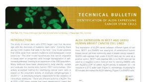 技术公告Identification of ALDH-Expressing Cancer Stem Cells Using ALDEFLUOR™
 挂图Small Molecules, Big Impact in Cancer Research Overview of signaling pathways and small molecules in cancer research


 沪公网安备31010102008431号