Sulforaphane targets pancreatic tumour-initiating cells by NF-kappaB-induced antiapoptotic signalling.
BACKGROUND AND AIMS: Emerging evidence suggests that highly treatment-resistant tumour-initiating cells (TICs) play a central role in the pathogenesis of pancreatic cancer. Tumour necrosis factor-related apoptosis-inducing ligand (TRAIL) is considered to be a novel anticancer agent; however,recent studies have shown that many pancreatic cancer cells are resistant to apoptosis induction by TRAIL due to TRAIL-activated nuclear factor-kappaB (NF-kappaB) signalling. Several chemopreventive agents are able to inhibit NF-kappaB,and favourable results have been obtained--for example,for the broccoli compound sulforaphane-in preventing metastasis in clinical studies. The aim of the study was to identify TICs in pancreatic carcinoma for analysis of resistance mechanisms and for definition of sensitising agents. METHODS: TICs were defined by expression patterns of a CD44(+)/CD24(-),CD44(+)/CD24(+) or CD44(+)/CD133(+) phenotype and correlation to growth in immunodeficient mice,differentiation grade,clonogenic growth,sphere formation,aldehyde dehydrogenase (ALDH) activity and therapy resistance. RESULTS: Mechanistically,specific binding of transcriptionally active cRel-containing NF-kappaB complexes in TICs was observed. Sulforaphane prevented NF-kappaB binding,downregulated apoptosis inhibitors and induced apoptosis,together with prevention of clonogenicity. Gemcitabine,the chemopreventive agents resveratrol and wogonin,and the death ligand TRAIL were less effective. In a xenograft model,sulforaphane strongly blocked tumour growth and angiogenesis,while combination with TRAIL had an additive effect without obvious cytotoxicity in normal cells. Freshly isolated patient tumour cells expressing markers for TICs could be sensitised by sulforaphane for TRAIL-induced cytotoxicity. CONCLUSION: The data provide new insights into resistance mechanisms of TICs and suggest the combination of sulforaphane with TRAIL as a promising strategy for targeting of pancreatic TICs.
View Publication
产品号#:
01700
01705
05751
01702
产品名:
ALDEFLUOR™ 试剂盒
ALDEFLUOR™ DEAB试剂, 1.5 mM, 1 mL
NeuroCult™ NS-A 扩增试剂盒(人)
ALDEFLUOR™检测缓冲液
Zhu X et al. (JUL 2010)
Molecular cancer therapeutics 9 7 2131--41
Identification of internalizing human single-chain antibodies targeting brain tumor sphere cells.
Glioblastoma multiforme (GBM) is the most common and aggressive form of primary brain tumor for which there is no curative treatment to date. Resistance to conventional therapies and tumor recurrence pose major challenges to treatment and management of this disease,and therefore new therapeutic strategies need to be developed. Previous studies by other investigators have shown that a subpopulation of GBM cells can grow as neurosphere-like cells when cultured in restrictive medium and exhibits enhanced tumor-initiating ability and resistance to therapy. We report here the identification of internalizing human single-chain antibodies (scFv) targeting GBM tumor sphere cells. We selected a large naive phage antibody display library on the glycosylation-dependent CD133 epitope-positive subpopulation of GBM cells grown as tumor spheres and identified internalizing scFvs that target tumor sphere cells broadly,as well as scFvs that target the CD133-positive subpopulation. These scFvs were found to be efficiently internalized by GBM tumor sphere cells. One scFv GC4 inhibited self-renewal of GBM tumor sphere cells in vitro. We have further developed a full-length human IgG1 based on this scFv,and found that it potently inhibits proliferation of GBM tumor sphere cells and GBM cells grown in regular nonselective medium. Taken together,these results show that internalizing human scFvs targeting brain tumor sphere cells can be readily identified from a phage antibody display library,which could be useful for further development of novel therapies that target subpopulations of GBM cells to combat recurrence and resistance to treatment.
View Publication
产品号#:
05751
产品名:
NeuroCult™ NS-A 扩增试剂盒(人)
Keysar SB and Jimeno A (SEP 2010)
Molecular cancer therapeutics 9 9 2450--7
More than markers: biological significance of cancer stem cell-defining molecules.
Small populations within an increasing array of solid tumors,labeled cancer stem cells (CSC) or tumor-initiating cells (TIC),have the ability to differentiate,self-renew,and replicate the original tumor in vivo. To date,these cells have been distinguished from the bulk-tumor population by the expression pattern of cell-surface proteins (e.g.,CD24,CD44,CD133) and cellular activities,such as the efflux of Hoechst dye or aldehyde dehydrogenase activity. Recent data have shown that these markers are inducible by exposure to anticancer agents; this finding highlights not only the potential fluidity of the CSC compartment,but also the functionality of these markers. The involvement of CD44 in invasion,adhesion,and metastasis,or the role of CD24 in modulation of src,FAK,and GLI1 are examples of these relevant roles. Instead of looking solely at the marker expression in these populations,we hope to clarify the biologically significant roles these markers and activities play in tumor progression,metastases,and as possible targets for therapy.
View Publication
产品号#:
01700
01705
01702
产品名:
ALDEFLUOR™ 试剂盒
ALDEFLUOR™ DEAB试剂, 1.5 mM, 1 mL
ALDEFLUOR™检测缓冲液
Simõ et al. (AUG 2011)
Breast cancer research and treatment 129 1 23--35
Effects of estrogen on the proportion of stem cells in the breast.
There is increasing evidence that breast cancers contain tumor-initiating cells with stem cell properties. The importance of estrogen in the development of the mammary gland and in breast cancer is well known,but the influence of estrogen on the stem cell population has not been assessed. We show that estrogen reduces the proportion of stem cells in the normal human mammary gland and in breast cancer cells. The embryonic stem cell genes NANOG,OCT4,and SOX2 are expressed in normal breast stem cells and at higher levels in breast tumor cells and their expression decreases upon differentiation. Overexpression of each stem cell gene reduces estrogen receptor (ER) expression,and increases the number of stem cells and their capacity for invasion,properties associated with tumorigenesis and poor prognosis. These results indicate that estrogen reduces the size of the human breast stem cell pool and may provide an explanation for the better prognosis of ER-positive tumors.
View Publication
产品号#:
01700
01705
01702
产品名:
ALDEFLUOR™ 试剂盒
ALDEFLUOR™ DEAB试剂, 1.5 mM, 1 mL
ALDEFLUOR™检测缓冲液
Wu K et al. (JAN 2011)
The Journal of biological chemistry 286 3 2132--42
Cell fate determination factor Dachshund reprograms breast cancer stem cell function.
The cell fate determination factor Dachshund was cloned as a dominant inhibitor of the hyperactive epidermal growth factor receptor ellipse. The expression of Dachshund is lost in human breast cancer associated with poor prognosis. Breast tumor-initiating cells (TIC) may contribute to tumor progression and therapy resistance. Here,endogenous DACH1 was reduced in breast cancer cell lines with high expression of TIC markers and in patient samples of the basal breast cancer phenotype. Re-expression of DACH1 reduced new tumor formation in serial transplantations in vivo,reduced mammosphere formation,and reduced the proportion of CD44(high)/CD24(low) breast tumor cells. Conversely,lentiviral shRNA to DACH1 increased the breast (B)TIC population. Genome-wide expression studies of mammary tumors demonstrated DACH1 repressed a molecular signature associated with stem cells (SOX2,Nanog,and KLF4) and genome-wide ChIP-seq analysis identified DACH1 binding to the promoter of the Nanog,KLF4,and Lin28 genes. KLF4/c-Myc and Oct4/Sox2 antagonized DACH1 repression of BTIC. Mechanistic studies demonstrated DACH1 directly repressed the Nanog and Sox2 promoters via a conserved domain. Endogenous DACH1 regulates BTIC in vitro and in vivo.
View Publication
产品号#:
01700
01705
01702
产品名:
ALDEFLUOR™ 试剂盒
ALDEFLUOR™ DEAB试剂, 1.5 mM, 1 mL
ALDEFLUOR™检测缓冲液
Yang X et al. (NOV 2010)
Cancer research 70 22 9463--72
Double-negative feedback loop between reprogramming factor LIN28 and microRNA let-7 regulates aldehyde dehydrogenase 1-positive cancer stem cells.
A relatively rare aldehyde dehydrogenase 1 (ALDH1)-positive stem cell-like" subpopulation of tumor cells has the unique ability to initiate and perpetuate tumor growth; moreover�
View Publication
产品号#:
01700
01705
01702
产品名:
ALDEFLUOR™ 试剂盒
ALDEFLUOR™ DEAB试剂, 1.5 mM, 1 mL
ALDEFLUOR™检测缓冲液
Chen Y-W et al. (NOV 2010)
Molecular cancer therapeutics 9 11 2879--92
Cucurbitacin I suppressed stem-like property and enhanced radiation-induced apoptosis in head and neck squamous carcinoma--derived CD44(+)ALDH1(+) cells.
Head and neck squamous cell carcinoma (HNSCC) is a prevalent cancer worldwide. Signal transducers and activators of transcription 3 (STAT3) signaling is reported to promote tumor malignancy and recurrence in HNSCC. Cucurbitacins,triterpenoid derivatives,are strong STAT3 inhibitors with anticancer properties. Recent studies have shown aldehyde dehydrogenase 1 (ALDH1) to be a marker of cancer stem cells (CSC) in HNSCC. The aim of this study was to investigate the therapeutic effect of cucurbitacin I in HNSCC-derived CSCs. Using immunohistochemical analysis,we firstly showed that CD44,ALDH1,and phosphorylated STAT3 (p-STAT3) were higher in high-grade HNSCCs,and that triple positivity for CD44/ALDH1/p-STAT3 indicated a worse prognosis for HNSCC patients. Secondly,CD44(+)ALDH1(+) cells isolated from seven HNSCC patients showed greater tumorigenicity,radioresistance,and high expression of stemness (Bmi-1/Oct-4/Nanog) and epithelial-mesenchymal-transitional (Snail/Twist) genes as p-STAT3 level increased. Furthermore,we found that cucurbitacin I (JSI-124) can effectively inhibit the expression of p-STAT3 and capacities for tumorigenicity,sphere formation,and radioresistance in HNSCC-CD44(+)ALDH1(+). Notably,150 nmol/L cucurbitacin I effectively blocked STAT3 signaling and downstream survivin and Bcl-2 expression,and it induced apoptosis in HNSCC-CD44(+)ALDH1(+). Moreover,microarray data indicated that 100 nmol/L cucurbitacin I facilitated CD44(+)ALDH1(+) cells to differentiate into CD44�?�ALDH1�?� and enhanced the radiosensitivity of HNSCC-CD44(+)ALDH1(+). Xenotransplant experiments revealed that cucurbitacin I combined with radiotherapy significantly suppressed tumorigenesis and lung metastasis and further improved the survival rate in HNSCC-CD44(+)ALDH1(+)-transplanted immunocompromised mice. Taken together,our data show that cucurbitacin I,STAT3 inhibitor,reduces radioresistant,distant-metastatic,and CSC-like properties of HNSCC-CD44(+)ALDH1(+) cells. The potential of cucurbitacin I as a radiosensitizer should be verified in future anti-CSC therapy.
View Publication
产品号#:
01700
01705
01702
产品名:
ALDEFLUOR™ 试剂盒
ALDEFLUOR™ DEAB试剂, 1.5 mM, 1 mL
ALDEFLUOR™检测缓冲液
Arbab AS et al. (SEP 2008)
FASEB journal : official publication of the Federation of American Societies for Experimental Biology 22 9 3234--46
Detection of migration of locally implanted AC133+ stem cells by cellular magnetic resonance imaging with histological findings.
This study investigated the factors responsible for migration and homing of magnetically labeled AC133(+) cells at the sites of active angiogenesis in tumor. AC133(+) cells labeled with ferumoxide-protamine sulfate were mixed with either rat glioma or human melanoma cells and implanted in flank of nude mice. An MRI of the tumors including surrounding tissues was performed. Tumor sections were stained for Prussian blue (PB),platelet-derived growth factor (PDGF),hypoxia-inducible factor-1alpha (HIF-1alpha),stromal cell derived factor-1 (SDF-1),matrix metalloproteinase-2 (MMP-2),vascular endothelial growth factor (VEGF),and endothelial markers. Fresh snap-frozen strips from the central and peripheral parts of the tumor were collected for Western blotting. MRIs demonstrated hypointense regions at the periphery of the tumors where the PB(+)/AC133(+) cells were positive for endothelial cells markers. At the sites of PB(+)/AC133(+) cells,both HIF-1alpha and SDF-1 were strongly positive and PDGF and MMP-2 showed generalized expression in the tumor and surrounding tissues. There was no significant association of PB(+)/AC133(+) cell localization and VEGF expression in tumor cells. Western blot demonstrated strong expression of the SDF-1,MMP-2,and PDGF at the peripheral parts of the tumors. HIF-1alpha was expressed at both the periphery and central parts of the tumor. This work demonstrates that magnetically labeled cells can be used as probes for MRI and histological identification of administered cells.
View Publication
GRP-78 secreted by tumor cells blocks the antiangiogenic activity of bortezomib.
Antiangiogenic effects of the proteasome inhibitor bortezomib were analyzed on tumor xenografts in vivo. Bortezomib strongly inhibited angiogenesis and vascularization in the chicken chorioallantoic membrane. Bortezomib's inhibitory effects on chorioallantoic membrane vascularization were abrogated in the presence of distinct tumor xenografts,thanks to a soluble factor secreted by tumor cells. Through size-exclusion and ion-exchange chromatography as well as mass spectroscopy,we identified GRP-78,a chaperone protein of the unfolded protein response,as being responsible for bortezomib resistance. Indeed,a variety of bortezomib-resistant solid tumor cell lines (PC-3,HRT-18),but not myeloma cell lines (U266,OPM-2),were able to secrete high amounts of GRP-78. Recombinant GRP-78 conferred bortezomib resistance to endothelial cells and OPM-2 myeloma cells. Knockdown of GRP78 gene expression in tumor cells and immunodepletion of GRP-78 protein from tumor cell supernatants restored bortezomib sensitivity. GRP-78 did not bind or complex bortezomib but induced prosurvival signals by phosphorylation of extracellular signal-related kinase and inhibited p53-mediated expression of proapoptotic Bok and Noxa proteins in endothelial cells. From our data,we conclude that distinct solid tumor cells are able to secrete GRP-78 into the tumor microenvironment,thus demonstrating a hitherto unknown mechanism of resistance to bortezomib.
View Publication
产品号#:
03814
产品名:
ClonaCell™-TCS 培养基
Su Y et al. (FEB 2010)
Cancer epidemiology,biomarkers & prevention : a publication of the American Association for Cancer Research,cosponsored by the American Society of Preventive Oncology 19 2 327--37
Aldehyde dehydrogenase 1 A1-positive cell population is enriched in tumor-initiating cells and associated with progression of bladder cancer.
Aldehyde dehydrogenase 1 A1 (ALDH1A1) has recently been suggested as a marker for cancer stem or stem-like cancer cells of some human malignancies. The purpose of this study was to investigate the stem cell-related function and clinical significance of the ALDH1A1 in bladder urothelial cell carcinoma. Aldefluor assay was used to isolate ALDH1A1+ cells from bladder cancer cells. Stem cell characteristics of the ALDH1A1+ cells were then investigated by in vitro and in vivo approaches. Immunohistochemistry was done for evaluating ALDH1A1 expression on 22 normal bladder tissues and 216 bladder tumor specimens of different stage and grade. The ALDH1A1+ cancer cells displayed higher in vitro tumorigenicity compared with isogenic ALDH1A1- cells. The ALDH1A1+ cancer cells could generate xenograft tumors that resembled the histopathologic characteristics and heterogeneity of the parental cells. High ALDH1A1 expression was found in 26% (56 of 216) of human bladder tumor specimens and significantly related to advanced pathologic stage,high histologic grade,recurrence and progression,and metastasis of bladder urothelial cell carcinomas (all P textless 0.05). Furthermore,ALDH1A1 expression was inversely associated with cancer-specific and overall survivals of the patients (P = 0.027 and 0.030,respectively). Therefore,ALDH1A1+ cell population could be enriched in tumor-initiating cells. ALDH1A1 may serve as a useful marker for monitoring the progression of bladder tumor and identifying bladder cancer patients with poor prognosis who might benefit from adjuvant and effective treatments.
View Publication

 EasySep™小鼠TIL(CD45)正选试剂盒


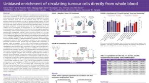 科学海报Unbiased Enrichment of Circulating Tumour Cells Directly from Whole Blood

 沪公网安备31010102008431号