A. Odawara et al. (JUL 2018)
Scientific reports 8 1 10416
Toxicological evaluation of convulsant and anticonvulsant drugs in human induced pluripotent stem cell-derived cortical neuronal networks using an MEA system.
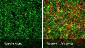
Functional evaluation assays using human induced pluripotent stem cell (hiPSC)-derived neurons can predict the convulsion toxicity of new drugs and the neurological effects of antiepileptic drugs. However,differences in responsiveness depending on convulsant type and antiepileptic drugs,and an evaluation index capable of comparing in vitro responses with in vivo responses are not well known. We observed the difference in synchronized burst patterns in the epileptiform activities induced by pentylentetrazole (PTZ) and 4-aminopryridine (4-AP) with different action mechanisms using multi-electrode arrays (MEAs); we also observed that 100 µM of the antiepileptic drug phenytoin suppressed epileptiform activities induced by PTZ,but increased those induced by 4-AP. To compare in vitro results with in vivo convulsive responses,frequency analysis of below 250 Hz,excluding the spike component,was performed. The in vivo convulsive firing enhancement of the high gamma$ wave and beta$ wave component were observed remarkably in in vitro hiPSC-derived neurons with astrocytes in co-culture. MEA measurement of hiPSC-derived neurons in co-culture with astrocytes and our analysis methods,including frequency analysis,appear effective for predicting convulsion toxicity,side effects,and their mechanism of action as well as the comparison of convulsions induced in vivo.
View Publication
产品号#:
05711
05790
05792
05793
05794
05795
100-1281
产品名:
NeuroCult™ SM1 神经添加物
BrainPhys™神经元培养基
BrainPhys™神经元培养基和SM1试剂盒
BrainPhys™ 神经元培养基N2-A和SM1试剂盒
BrainPhys™原代神经元试剂盒
BrainPhys™ hPSC 神经元试剂盒
NeuroCult™ SM1 神经添加物
Griesi-Oliveira K et al. (NOV 2014)
Molecular psychiatry 20 March 1--16
Modeling non-syndromic autism and the impact of TRPC6 disruption in human neurons.
An increasing number of genetic variants have been implicated in autism spectrum disorders (ASDs),and the functional study of such variants will be critical for the elucidation of autism pathophysiology. Here,we report a de novo balanced translocation disruption of TRPC6,a cation channel,in a non-syndromic autistic individual. Using multiple models,such as dental pulp cells,induced pluripotent stem cell (iPSC)-derived neuronal cells and mouse models,we demonstrate that TRPC6 reduction or haploinsufficiency leads to altered neuronal development,morphology and function. The observed neuronal phenotypes could then be rescued by TRPC6 complementation and by treatment with insulin-like growth factor-1 or hyperforin,a TRPC6-specific agonist,suggesting that ASD individuals with alterations in this pathway may benefit from these drugs. We also demonstrate that methyl CpG binding protein-2 (MeCP2) levels affect TRPC6 expression. Mutations in MeCP2 cause Rett syndrome,revealing common pathways among ASDs. Genetic sequencing of TRPC6 in 1041 ASD individuals and 2872 controls revealed significantly more nonsynonymous mutations in the ASD population,and identified loss-of-function mutations with incomplete penetrance in two patients. Taken together,these findings suggest that TRPC6 is a novel predisposing gene for ASD that may act in a multiple-hit model. This is the first study to use iPSC-derived human neurons to model non-syndromic ASD and illustrate the potential of modeling genetically complex sporadic diseases using such cells.Molecular Psychiatry advance online publication,11 November 2014; doi:10.1038/mp.2014.141.
View Publication
产品号#:
05850
05857
05870
05875
85850
85857
85870
85875
产品名:
mTeSR™1
mTeSR™1
Wang S et al. (MAR 2015)
Sci Rep 5 9232
Differentiation of human induced pluripotent stem cells to mature functional Purkinje neurons.
It remains a challenge to differentiate human induced pluripotent stem cells (iPSCs) or embryonic stem (ES) cells to Purkinje cells. In this study,we derived iPSCs from human fibroblasts and directed the specification of iPSCs first to Purkinje progenitors,by adding Fgf2 and insulin to the embryoid bodies (EBs) in a time-sensitive manner,which activates the endogenous production of Wnt1 and Fgf8 from EBs that further patterned the cells towards a midbrain-hindbrain-boundary tissue identity. Neph3-positive human Purkinje progenitors were sorted out by using flow cytometry and cultured either alone or with granule cell precursors,in a 2-dimensional or 3-dimensional environment. However,Purkinje progenitors failed to mature further under above conditions. By co-culturing human Purkinje progenitors with rat cerebellar slices,we observed mature Purkinje-like cells with right morphology and marker expression patterns,which yet showed no appropriate membrane properties. Co-culture with human fetal cerebellar slices drove the progenitors to not only morphologically correct but also electrophysiologically functional Purkinje neurons. Neph3-posotive human cells could also survive transplantation into the cerebellum of newborn immunodeficient mice and differentiate to L7- and Calbindin-positive neurons. Obtaining mature human Purkinje cells in vitro has significant implications in studying the mechanisms of spinocerebellar ataxias and other cerebellar diseases.
View Publication
产品号#:
05850
05857
05870
05875
85850
85857
85870
85875
产品名:
mTeSR™1
mTeSR™1
Carmona-Mora P et al. (OCT 2015)
Human Genetics 134 10 1099--1115
The nuclear localization pattern and interaction partners of GTF2IRD1 demonstrate a role in chromatin regulation
GTF2IRD1 is one of the three members of the GTF2I gene family,clustered on chromosome 7 within a 1.8 Mb region that is prone to duplications and deletions in humans. Hemizygous deletions cause Williams-Beuren syndrome (WBS) and duplications cause WBS duplication syndrome. These copy number variations disturb a variety of developmental systems and neurological functions. Human mapping data and analyses of knockout mice show that GTF2IRD1 and GTF2I underpin the craniofacial abnormalities,mental retardation,visuospatial deficits and hypersociability of WBS. However,the cellular role of the GTF2IRD1 protein is poorly understood due to its very low abundance and a paucity of reagents. Here,for the first time,we show that endogenous GTF2IRD1 has a punctate pattern in the nuclei of cultured human cell lines and neurons. To probe the functional relationships of GTF2IRD1 in an unbiased manner,yeast two-hybrid libraries were screened,isolating 38 novel interaction partners,which were validated in mammalian cell lines. These relationships illustrate GTF2IRD1 function,as the isolated partners are mostly involved in chromatin modification and transcriptional regulation,whilst others indicate an unexpected role in connection with the primary cilium. Mapping of the sites of protein interaction also indicates key features regarding the evolution of the GTF2IRD1 protein. These data provide a visual and molecular basis for GTF2IRD1 nuclear function that will lead to an understanding of its role in brain,behaviour and human disease.
View Publication
产品号#:
05850
05857
05870
05875
85850
85857
85870
85875
产品名:
mTeSR™1
mTeSR™1
Fuller HR et al. (JAN 2015)
Frontiers in cellular neuroscience 9 January 506
Spinal Muscular Atrophy Patient iPSC-Derived Motor Neurons Have Reduced Expression of Proteins Important in Neuronal Development.
Spinal muscular atrophy (SMA) is an inherited neuromuscular disease primarily characterized by degeneration of spinal motor neurons,and caused by reduced levels of the SMN protein. Previous studies to understand the proteomic consequences of reduced SMN have mostly utilized patient fibroblasts and animal models. We have derived human motor neurons from type I SMA and healthy controls by creating their induced pluripotent stem cells (iPSCs). Quantitative mass spectrometry of these cells revealed increased expression of 63 proteins in control motor neurons compared to respective fibroblasts,whereas 30 proteins were increased in SMA motor neurons vs. their fibroblasts. Notably,UBA1 was significantly decreased in SMA motor neurons,supporting evidence for ubiquitin pathway defects. Subcellular distribution of UBA1 was predominantly cytoplasmic in SMA motor neurons in contrast to nuclear in control motor neurons; suggestive of neurodevelopmental abnormalities. Many of the proteins that were decreased in SMA motor neurons,including beta III-tubulin and UCHL1,were associated with neurodevelopment and differentiation. These neuron-specific consequences of SMN depletion were not evident in fibroblasts,highlighting the importance of iPSC technology. The proteomic profiles identified here provide a useful resource to explore the molecular consequences of reduced SMN in motor neurons,and for the identification of novel biomarker and therapeutic targets for SMA.
View Publication
产品号#:
05832
05850
05857
05870
05875
85850
85857
85870
85875
产品名:
STEMdiff™ 神经花环选择试剂
mTeSR™1
mTeSR™1
Chamma I et al. (MAR 2016)
Nature Communications 7 10773
Mapping the dynamics and nanoscale organization of synaptic adhesion proteins using monomeric streptavidin
The advent of super-resolution imaging (SRI) has created a need for optimized labelling strategies. We present a new method relying on fluorophore-conjugated monomeric streptavidin (mSA) to label membrane proteins carrying a short,enzymatically biotinylated tag,compatible with SRI techniques including uPAINT,STED and dSTORM. We demonstrate efficient and specific labelling of target proteins in confined intercellular and organotypic tissues,with reduced steric hindrance and no crosslinking compared with multivalent probes. We use mSA to decipher the dynamics and nanoscale organization of the synaptic adhesion molecules neurexin-1β,neuroligin-1 (Nlg1) and leucine-rich-repeat transmembrane protein 2 (LRRTM2) in a dual-colour configuration with GFP nanobody,and show that these proteins are diffusionally trapped at synapses where they form apposed trans-synaptic adhesive structures. Furthermore,Nlg1 is dynamic,disperse and sensitive to synaptic stimulation,whereas LRRTM2 is organized in compact and stable nanodomains. Thus,mSA is a versatile tool to image membrane proteins at high resolution in complex live environments,providing novel information about the nano-organization of biological structures.
View Publication
产品号#:
05711
100-1281
产品名:
NeuroCult™ SM1 神经添加物
NeuroCult™ SM1 神经添加物
Kim YY et al. (SEP 2016)
PLOS ONE 11 9 e0163812
Alcohol-Induced Molecular Dysregulation in Human Embryonic Stem Cell-Derived Neural Precursor Cells
Adverse effect of alcohol on neural function has been well documented. Especially,the teratogenic effect of alcohol on neurodevelopment during embryogenesis has been demonstrated in various models,which could be a pathologic basis for fetal alcohol spectrum disorders (FASDs). While the developmental defects from alcohol abuse during gestation have been described,the specific mechanisms by which alcohol mediates these injuries have yet to be determined. Recent studies have shown that alcohol has significant effect on molecular and cellular regulatory mechanisms in embryonic stem cell (ESC) differentiation including genes involved in neural development. To test our hypothesis that alcohol induces molecular alterations during neural differentiation we have derived neural precursor cells from pluripotent human ESCs in the presence or absence of ethanol treatment. Genome-wide transcriptomic profiling identified molecular alterations induced by ethanol exposure during neural differentiation of hESCs into neural rosettes and neural precursor cell populations. The Database for Annotation,Visualization and Integrated Discovery (DAVID) functional analysis on significantly altered genes showed potential ethanol's effect on JAK-STAT signaling pathway,neuroactive ligand-receptor interaction,Toll-like receptor (TLR) signaling pathway,cytokine-cytokine receptor interaction and regulation of autophagy. We have further quantitatively verified ethanol-induced alterations of selected candidate genes. Among verified genes we further examined the expression of P2RX3,which is associated with nociception,a peripheral pain response. We found ethanol significantly reduced the level of P2RX3 in undifferentiated hESCs,but induced the level of P2RX3 mRNA and protein in hESC-derived NPCs. Our result suggests ethanol-induced dysregulation of P2RX3 along with alterations in molecules involved in neural activity such as neuroactive ligand-receptor interaction may be a molecular event associated with alcohol-related peripheral neuropathy of an enhanced nociceptive response.
View Publication
Yanpallewar SU et al. (JAN 2010)
The Journal of neuroscience : the official journal of the Society for Neuroscience 30 3 1096--109
Alpha2-adrenoceptor blockade accelerates the neurogenic, neurotrophic, and behavioral effects of chronic antidepressant treatment.
Slow-onset adaptive changes that arise from sustained antidepressant treatment,such as enhanced adult hippocampal neurogenesis and increased trophic factor expression,play a key role in the behavioral effects of antidepressants. alpha(2)-Adrenoceptors contribute to the modulation of mood and are potential targets for the development of faster acting antidepressants. We investigated the influence of alpha(2)-adrenoceptors on adult hippocampal neurogenesis. Our results indicate that alpha(2)-adrenoceptor agonists,clonidine and guanabenz,decrease adult hippocampal neurogenesis through a selective effect on the proliferation,but not the survival or differentiation,of progenitors. These effects persist in dopamine beta-hydroxylase knock-out (Dbh(-/-)) mice lacking norepinephrine,supporting a role for alpha(2)-heteroceptors on progenitor cells,rather than alpha(2)-autoreceptors on noradrenergic neurons that inhibit norepinephrine release. Adult hippocampal progenitors in vitro express all the alpha(2)-adrenoceptor subtypes,and decreased neurosphere frequency and BrdU incorporation indicate direct effects of alpha(2)-adrenoceptor stimulation on progenitors. Furthermore,coadministration of the alpha(2)-adrenoceptor antagonist yohimbine with the antidepressant imipramine significantly accelerates effects on hippocampal progenitor proliferation,the morphological maturation of newborn neurons,and the increase in expression of brain derived neurotrophic factor and vascular endothelial growth factor implicated in the neurogenic and behavioral effects of antidepressants. Finally,short-duration (7 d) yohimbine and imipramine treatment results in robust behavioral responses in the novelty suppressed feeding test,which normally requires 3 weeks of treatment with classical antidepressants. Our results demonstrate that alpha(2)-adrenoceptors,expressed by progenitor cells,decrease adult hippocampal neurogenesis,while their blockade speeds up antidepressant action,highlighting their importance as targets for faster acting antidepressants.
View Publication
NMDA receptors mediate synaptic competition in culture.
BACKGROUND: Activity through NMDA type glutamate receptors sculpts connectivity in the developing nervous system. This topic is typically studied in the visual system in vivo,where activity of inputs can be differentially regulated,but in which individual synapses are difficult to visualize and mechanisms governing synaptic competition can be difficult to ascertain. Here,we develop a model of NMDA-receptor dependent synaptic competition in dissociated cultured hippocampal neurons. METHODOLOGY/PRINCIPAL FINDINGS: GluN1 -/- (KO) mouse hippocampal neurons lacking the essential NMDA receptor subunit were cultured alone or cultured in defined ratios with wild type (WT) neurons. The absence of functional NMDA receptors did not alter neuron survival. Synapse development was assessed by immunofluorescence for postsynaptic PSD-95 family scaffold and apposed presynaptic vesicular glutamate transporter VGlut1. Synapse density was specifically enhanced onto minority wild type neurons co-cultured with a majority of GluN1 -/- neighbour neurons,both relative to the GluN1 -/- neighbours and relative to sister pure wild type cultures. This form of synaptic competition was dependent on NMDA receptor activity and not conferred by the mere physical presence of GluN1. In contrast to these results in 10% WT and 90% KO co-cultures,synapse density did not differ by genotype in 50% WT and 50% KO co-cultures or in 90% WT and 10% KO co-cultures. CONCLUSIONS/SIGNIFICANCE: The enhanced synaptic density onto NMDA receptor-competent neurons in minority coculture with GluN1 -/- neurons represents a cell culture paradigm for studying synaptic competition. Mechanisms involved may include a retrograde 'reward' signal generated by WT neurons,although in this paradigm there was no 'punishment' signal against GluN1 -/- neurons. Cell culture assays involving such defined circuits may help uncover the rules and mechanisms of activity-dependent synaptic competition in the developing nervous system.
View Publication

 EasySep™小鼠TIL(CD45)正选试剂盒


 科学海报Single-Cell RNA Sequencing Analysis of Regionally Patterned Human Pluripotent Stem Cell-Derived Neural Organoids
 沪公网安备31010102008431号