Katori S et al. (JUL 2009)
The Journal of neuroscience : the official journal of the Society for Neuroscience 29 29 9137--47
Protocadherin-alpha family is required for serotonergic projections to appropriately innervate target brain areas.
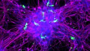
Serotonergic axons from the raphe nuclei in the brainstem project to every region of the brain,where they make connections through their extensive terminal arborizations. This serotonergic innervation contributes to various normal behaviors and psychiatric disorders. The protocadherin-alpha (Pcdha) family of clustered protocadherins consists of 14 cadherin-related molecules generated from a single gene cluster. We found that the Pcdhas were strongly expressed in the serotonergic neurons. To elucidate their roles,we examined serotonergic fibers in a mouse mutant (Pcdha(Delta CR/Delta CR)) lacking the Pcdha cytoplasmic region-encoding exons,which are common to the gene cluster. In the first week after birth,the distribution pattern of serotonergic fibers in Pcdha(Delta CR/Delta CR) mice was similar to wild-type,but by 3 weeks of age,when the serotonergic axonal termini complete their arborizations,the distribution of the projections was abnormal. In some target regions,notably the globus pallidus and substantia nigra,the normally even distribution of serotonin axonal terminals was,in the mutants,dense at the periphery of each region,but sparse in the center. In the stratum lacunosum-molecular of the hippocampus,the mutants showed denser serotonergic innervation than in wild-type,and in the dentate gyrus of the hippocampus and the caudate-putamen,the innervation was sparser. Together,the abnormalities suggested that Pcdha proteins are important in the late-stage maturation of serotonergic projections. Further examination of alternatively spliced exons encoding the cytoplasmic tail showed that the A-type (but not the B-type) cytoplasmic tail was essential for the normal development of serotonergic projections.
View Publication
Dobie FA and Craig AM (JUL 2011)
The Journal of neuroscience : the official journal of the Society for Neuroscience 31 29 10481--93
Inhibitory synapse dynamics: coordinated presynaptic and postsynaptic mobility and the major contribution of recycled vesicles to new synapse formation.
Dynamics of GABAergic synaptic components have been studied previously over milliseconds to minutes,revealing mobility of postsynaptic scaffolds and receptors. Here we image inhibitory synapses containing fluorescently tagged postsynaptic scaffold Gephyrin,together with presynaptic vesicular GABA transporter (VGAT) or postsynaptic GABA(A) receptor γ2 subunit (GABA(A)Rγ2),over seconds to days in cultured rat hippocampal neurons,revealing modes of inhibitory synapse formation and remodeling. Entire synapses were mobile,translocating rapidly within a confined region and exhibiting greater nonstochastic motion over multihour periods. Presynaptic and postsynaptic components moved in unison,maintaining close apposition while translocating distances of several micrometers. An observed flux in the density of synaptic puncta partially resulted from the apparent merging and splitting of preexisting clusters. De novo formation of inhibitory synapses was observed,marked by the appearance of stably apposed Gephyrin and VGAT clusters at sites previously lacking either component. Coclustering of GABA(A)Rγ2 supports the identification of such new clusters as synapses. Nascent synapse formation occurred by gradual accumulation of components over several hours,with VGAT clustering preceding that of Gephyrin and GABA(A)Rγ2. Comparing VGAT labeling by active uptake of a luminal domain antibody with post hoc immunocytochemistry indicated that recycling vesicles from preexisting boutons significantly contribute to vesicle pools at the majority of new inhibitory synapses. Although new synapses formed primarily on dendrite shafts,some also formed on dendritic protrusions,without apparent interconversion. Altogether,the long-term imaging of GABAergic presynaptic and postsynaptic components reveals complex dynamics and perpetual remodeling with implications for mechanisms of assembly and synaptic integration.
View Publication
产品号#:
05711
100-1281
产品名:
NeuroCult™ SM1 神经添加物
NeuroCult™ SM1 神经添加物
Pecho-Vrieseling E et al. (AUG 2014)
Nat Neurosci 17 8 1064--1072
Transneuronal propagation of mutant huntingtin contributes to non-cell autonomous pathology in neurons.
In Huntington's disease (HD),whether transneuronal spreading of mutant huntingtin (mHTT) occurs and its contribution to non-cell autonomous damage in brain networks is largely unknown. We found mHTT spreading in three different neural network models: human neurons integrated in the neural network of organotypic brain slices of HD mouse model,an ex vivo corticostriatal slice model and the corticostriatal pathway in vivo. Transneuronal propagation of mHTT was blocked by two different botulinum neurotoxins,each known for specifically inactivating a single critical component of the synaptic vesicle fusion machinery. Moreover,healthy human neurons in HD mouse model brain slices displayed non-cell autonomous changes in morphological integrity that were more pronounced when these neurons bore mHTT aggregates. Altogether,our findings suggest that transneuronal propagation of mHTT might be an important and underestimated contributor to the pathophysiology of HD.
View Publication
产品号#:
05850
05857
05870
05875
85850
85857
85870
85875
产品名:
mTeSR™1
mTeSR™1
Chen C et al. (JUL 2014)
Nature communications 5 4430
Role of astroglia in Down's syndrome revealed by patient-derived human-induced pluripotent stem cells.
Down's syndrome (DS),caused by trisomy of human chromosome 21,is the most common genetic cause of intellectual disability. Here we use induced pluripotent stem cells (iPSCs) derived from DS patients to identify a role for astrocytes in DS pathogenesis. DS astroglia exhibit higher levels of reactive oxygen species and lower levels of synaptogenic molecules. Astrocyte-conditioned medium collected from DS astroglia causes toxicity to neurons,and fails to promote neuronal ion channel maturation and synapse formation. Transplantation studies show that DS astroglia do not promote neurogenesis of endogenous neural stem cells in vivo. We also observed abnormal gene expression profiles from DS astroglia. Finally,we show that the FDA-approved antibiotic drug,minocycline,partially corrects the pathological phenotypes of DS astroglia by specifically modulating the expression of S100B,GFAP,inducible nitric oxide synthase,and thrombospondins 1 and 2 in DS astroglia. Our studies shed light on the pathogenesis and possible treatment of DS by targeting astrocytes with a clinically available drug.
View Publication
K. B. Langer et al. (APR 2018)
Stem cell reports 10 4 1282--1293
Retinal Ganglion Cell Diversity and Subtype Specification from Human Pluripotent Stem Cells.
Retinal ganglion cells (RGCs) are the projection neurons of the retina and transmit visual information to postsynaptic targets in the brain. While this function is shared among nearly all RGCs,this class of cell is remarkably diverse,comprised of multiple subtypes. Previous efforts have identified numerous RGC subtypes in animal models,but less attention has been paid to human RGCs. Thus,efforts of this study examined the diversity of RGCs differentiated from human pluripotent stem cells (hPSCs) and characterized defined subtypes through the expression of subtype-specific markers. Further investigation of these subtypes was achieved using single-cell transcriptomics,confirming the combinatorial expression of molecular markers associated with these subtypes,and also provided insight into more subtype-specific markers. Thus,the results of this study describe the derivation of RGC subtypes from hPSCs and will support the future exploration of phenotypic and functional diversity within human RGCs.
View Publication
产品号#:
05790
05792
05793
05794
05795
85850
85857
产品名:
BrainPhys™神经元培养基
BrainPhys™神经元培养基和SM1试剂盒
BrainPhys™ 神经元培养基N2-A和SM1试剂盒
BrainPhys™原代神经元试剂盒
BrainPhys™ hPSC 神经元试剂盒
mTeSR™1
mTeSR™1
A. M. Tukker et al. (JUL 2018)
Neurotoxicology 67 215--225
Human iPSC-derived neuronal models for in vitro neurotoxicity assessment.
Neurotoxicity testing still relies on ethically debated,expensive and time consuming in vivo experiments,which are unsuitable for high-throughput toxicity screening. There is thus a clear need for a rapid in vitro screening strategy that is preferably based on human-derived neurons to circumvent interspecies translation. Recent availability of commercially obtainable human induced pluripotent stem cell (hiPSC)-derived neurons and astrocytes holds great promise in assisting the transition from the current standard of rat primary cortical cultures to an animal-free alternative. We therefore composed several hiPSC-derived neuronal models with different ratios of excitatory and inhibitory neurons in the presence or absence of astrocytes. Using immunofluorescent stainings and multi-well micro-electrode array (mwMEA) recordings we demonstrate that these models form functional neuronal networks that become spontaneously active. The differences in development of spontaneous neuronal activity and bursting behavior as well as spiking patterns between our models confirm the importance of the presence of astrocytes. Preliminary neurotoxicity assessment demonstrates that these cultures can be modulated with known seizurogenic compounds,such as picrotoxin (PTX) and endosulfan,and the neurotoxicant methylmercury (MeHg). However,the chemical-induced effects on different parameters for neuronal activity,such as mean spike rate (MSR) and mean burst rate (MBR),may depend on the ratio of inhibitory and excitatory neurons. Our results thus indicate that hiPSC-derived neuronal models must be carefully designed and characterized prior to large-scale use in neurotoxicity screening.
View Publication

 EasySep™小鼠TIL(CD45)正选试剂盒


 实验方案How to Co-Culture Human Pluripotent Stem Cell (hPSC)-Derived Forebrain Neurons and Microglia
 实验方案How to Dissociate 3D Neural Organoids into a Single-Cell Suspension

 沪公网安备31010102008431号