Tagliafierro L et al. (NOV 2017)
Alzheimer's & dementia : the journal of the Alzheimer's Association 13 11 1237--1250
Genetic analysis of α-synuclein 3' untranslated region and its corresponding microRNAs in relation to Parkinson's disease compared to dementia with Lewy bodies.
INTRODUCTION The α-synuclein (SNCA) gene has been implicated in the etiology of Parkinson's disease (PD) and dementia with Lewy bodies (DLB). METHODS A computational analysis of SNCA 3' untranslated region to identify potential microRNA (miRNA) binding sites and quantitative real-time polymerase chain reaction (PCR) to determine their expression in isogenic induced pluripotent stem cell-derived dopaminergic and cholinergic neurons as a model of PD and DLB,respectively,were performed. In addition,we performed a deep sequencing analysis of the SNCA 3' untranslated region of autopsy-confirmed cases of PD,DLB,and normal controls,followed by genetic association analysis of the identified variants. RESULTS We identified four miRNA binding sites and observed a neuronal-type-specific expression profile for each miRNA in the different isogenic induced pluripotent stem cell-derived dopaminergic and cholinergic neurons. Furthermore,we found that the short structural variant rs777296100-polyT was moderately associated with DLB but not with PD. DISCUSSION We suggest that the regulation of SNCA expression through miRNAs is neuronal-type-specific and possibly plays a part in the phenotypic heterogeneity of synucleinopathies. Furthermore,genetic variability in the SNCA gene may contribute to synucleinopathies in a pathology-specific manner.
View Publication
产品号#:
05790
05792
05793
05794
05795
产品名:
BrainPhys™神经元培养基
BrainPhys™神经元培养基和SM1试剂盒
BrainPhys™ 神经元培养基N2-A和SM1试剂盒
BrainPhys™原代神经元试剂盒
BrainPhys™ hPSC 神经元试剂盒
Busskamp V et al. (NOV 2014)
Molecular systems biology 10 11 760
Rapid neurogenesis through transcriptional activation in human stem cells.
Advances in cellular reprogramming and stem cell differentiation now enable ex vivo studies of human neuronal differentiation. However,it remains challenging to elucidate the underlying regulatory programs because differentiation protocols are laborious and often result in low neuron yields. Here,we overexpressed two Neurogenin transcription factors in human-induced pluripotent stem cells and obtained neurons with bipolar morphology in 4 days,at greater than 90% purity. The high purity enabled mRNA and microRNA expression profiling during neurogenesis,thus revealing the genetic programs involved in the rapid transition from stem cell to neuron. The resulting cells exhibited transcriptional,morphological and functional signatures of differentiated neurons,with greatest transcriptional similarity to prenatal human brain samples. Our analysis revealed a network of key transcription factors and microRNAs that promoted loss of pluripotency and rapid neurogenesis via progenitor states. Perturbations of key transcription factors affected homogeneity and phenotypic properties of the resulting neurons,suggesting that a systems-level view of the molecular biology of differentiation may guide subsequent manipulation of human stem cells to rapidly obtain diverse neuronal types.
View Publication
产品号#:
05854
05855
05850
05857
05870
05875
85850
85857
85870
85875
产品名:
mFreSR™
mFreSR™
mTeSR™1
mTeSR™1
Wattanapanitch M et al. (SEP 2014)
PloS one 9 9 e106952
Dual small-molecule targeting of SMAD signaling stimulates human induced pluripotent stem cells toward neural lineages.
Incurable neurological disorders such as Parkinson's disease (PD),Huntington's disease (HD),and Alzheimer's disease (AD) are very common and can be life-threatening because of their progressive disease symptoms with limited treatment options. To provide an alternative renewable cell source for cell-based transplantation and as study models for neurological diseases,we generated induced pluripotent stem cells (iPSCs) from human dermal fibroblasts (HDFs) and then differentiated them into neural progenitor cells (NPCs) and mature neurons by dual SMAD signaling inhibitors. Reprogramming efficiency was improved by supplementing the histone deacethylase inhibitor,valproic acid (VPA),and inhibitor of p160-Rho associated coiled-coil kinase (ROCK),Y-27632,after retroviral transduction. We obtained a number of iPS colonies that shared similar characteristics with human embryonic stem cells in terms of their morphology,cell surface antigens,pluripotency-associated gene and protein expressions as well as their in vitro and in vivo differentiation potentials. After treatment with Noggin and SB431542,inhibitors of the SMAD signaling pathway,HDF-iPSCs demonstrated rapid and efficient differentiation into neural lineages. Six days after neural induction,neuroepithelial cells (NEPCs) were observed in the adherent monolayer culture,which had the ability to differentiate further into NPCs and neurons,as characterized by their morphology and the expression of neuron-specific transcripts and proteins. We propose that our study may be applied to generate neurological disease patient-specific iPSCs allowing better understanding of disease pathogenesis and drug sensitivity assays.
View Publication
Lippmann ES et al. (APR 2014)
Stem Cells 32 4 1032--1042
Defined human pluripotent stem cell culture enables highly efficient neuroepithelium derivation without small molecule inhibitors.
The embryonic neuroepithelium gives rise to the entire central nervous system in vivo,making it an important tissue for developmental studies and a prospective cell source for regenerative applications. Current protocols for deriving homogenous neuroepithelial cultures from human pluripotent stem cells (hPSCs) consist of either embryoid body-mediated neuralization followed by a manual isolation step or adherent differentiation using small molecule inhibitors. Here,we report that hPSCs maintained under chemically defined,feeder-independent,and xeno-free conditions can be directly differentiated into pure neuroepithelial cultures ([mt]90% Pax6(+)/N-cadherin(+) with widespread rosette formation) within 6 days under adherent conditions,without small molecule inhibitors,and using only minimalistic medium consisting of Dulbecco's modified Eagle's medium/F-12,sodium bicarbonate,selenium,ascorbic acid,transferrin,and insulin (i.e.,E6 medium). Furthermore,we provide evidence that the defined culture conditions enable this high level of neural conversion in contrast to hPSCs maintained on mouse embryonic fibroblasts (MEFs). In addition,hPSCs previously maintained on MEFs could be rapidly converted to a neural compliant state upon transfer to these defined conditions while still maintaining their ability to generate all three germ layers. Overall,this fully defined and scalable protocol should be broadly useful for generating therapeutic neural cells for regenerative applications.
View Publication
Yamazaki K et al. (DEC 2016)
Journal of Biomolecular Screening 21 10 1054--1064
Functional Comparison of Neuronal Cells Differentiated from Human Induced Pluripotent Stem CellDerived Neural Stem Cells under Different Oxygen and Medium Conditions
Because neurons are difficult to obtain from humans,generating functional neurons from human induced pluripotent stem cells (hiPSCs) is important for establishing physiological or disease-relevant screening systems for drug discovery. To examine the culture conditions leading to efficient differentiation of functional neural cells,we investigated the effects of oxygen stress (2% or 20% O2) and differentiation medium (DMEM/F12:Neurobasal-based [DN] or commercial [PhoenixSongs Biologicals; PS]) on the expression of genes related to neural differentiation,glutamate receptor function,and the formation of networks of neurons differentiated from hiPSCs (201B7) via long-term self-renewing neuroepithelial-like stem (lt-NES) cells. Expression of genes related to neural differentiation occurred more quickly in PS and/or 2% O2 than in DN and/or 20% O2,resulting in high responsiveness of neural cells to glutamate,N-methyl-d-aspartate (NMDA),α-amino-3-hydroxy-5-methyl-4-isoxazolepropionate (AMPA),and (S)-3,5-d...
View Publication
产品号#:
05832
产品名:
STEMdiff™ 神经花环选择试剂
C. L. Moreno et al. ( 2018)
Molecular neurodegeneration 13 1 33
BACKGROUND Type 2 diabetes (T2D) is a recognized risk factor for the development of cognitive impairment (CI) and/or dementia,although the exact nature of the molecular pathology of T2D-associated CI remains obscure. One link between T2D and CI might involve decreased insulin signaling in brain and/or neurons in either animal or postmortem human brains as has been reported as a feature of Alzheimer's disease (AD). Here we asked if neuronal insulin resistance is a cell autonomous phenomenon in a familial form of AD. METHODS We have applied a newly developed protocol for deriving human basal forebrain cholinergic neurons (BFCN) from skin fibroblasts via induced pluripotent stem cell (iPSC) technology. We generated wildtype and familial AD mutant PSEN2 N141I (presenilin 2) BFCNs and assessed if insulin signaling,insulin regulation of the major AD proteins Abeta$ and/or tau,and/or calcium fluxes is altered by the PSEN2 N141I mutation. RESULTS We report herein that wildtype,PSEN2 N141I and CRISPR/Cas9-corrected iPSC-derived BFCNs (and their precursors) show indistinguishable insulin signaling profiles as determined by the phosphorylation of canonical insulin signaling pathway molecules. Chronic insulin treatment of BFCNs of all genotypes led to a reduction in the Abeta$42/40 ratio. Unexpectedly,we found a CRISPR/Cas9-correctable effect of PSEN2 N141I on calcium flux,which could be prevented by chronic exposure of BFCNs to insulin. CONCLUSIONS Our studies indicate that the familial AD mutation PSEN2 N141I does not induce neuronal insulin resistance in a cell autonomous fashion. The ability of insulin to correct calcium fluxes and to lower Abeta$42/40 ratio suggests that insulin acts to oppose an AD-pathophysiology. Hence,our results are consistent with a potential physiological role for insulin as a mediator of resilience by counteracting specific metabolic and molecular features of AD.
View Publication
产品号#:
07920
07922
05790
05792
05793
05794
05795
85850
85857
85870
85875
05791
产品名:
ACCUTASE™
ACCUTASE™
BrainPhys™神经元培养基
BrainPhys™神经元培养基和SM1试剂盒
BrainPhys™ 神经元培养基N2-A和SM1试剂盒
BrainPhys™原代神经元试剂盒
BrainPhys™ hPSC 神经元试剂盒
mTeSR™1
mTeSR™1
BrainPhys™ 无酚红
P. H. Chia et al. (MAY 2018)
eLife 7
A homozygous loss-of-function CAMK2A mutation causes growth delay, frequent seizures and severe intellectual disability.
Calcium/calmodulin-dependent protein kinase II (CAMK2) plays fundamental roles in synaptic plasticity that underlies learning and memory. Here,we describe a new recessive neurodevelopmental syndrome with global developmental delay,seizures and intellectual disability. Using linkage analysis and exome sequencing,we found that this disease maps to chromosome 5q31.1-q34 and is caused by a biallelic germline mutation in CAMK2A. The missense mutation,p.His477Tyr is located in the CAMK2A association domain that is critical for its function and localization. Biochemically,the p.His477Tyr mutant is defective in self-oligomerization and unable to assemble into the multimeric holoenzyme.In vivo,CAMK2AH477Y failed to rescue neuronal defects in C. elegans lacking unc-43,the ortholog of human CAMK2A. In vitro,neurons derived from patient iPSCs displayed profound synaptic defects. Together,our data demonstrate that a recessive germline mutation in CAMK2A leads to neurodevelopmental defects in humans and suggest that dysfunctional CAMK2 paralogs may contribute to other neurological disorders.
View Publication
产品号#:
05790
05792
05793
05794
05795
85850
85857
85870
85875
产品名:
BrainPhys™神经元培养基
BrainPhys™神经元培养基和SM1试剂盒
BrainPhys™ 神经元培养基N2-A和SM1试剂盒
BrainPhys™原代神经元试剂盒
BrainPhys™ hPSC 神经元试剂盒
mTeSR™1
mTeSR™1
Basma H et al. (MAR 2014)
American journal of physiology. Lung cellular and molecular physiology 306 6 L552--65
Reprogramming of COPD lung fibroblasts through formation of induced pluripotent stem cells.
Reprogramming somatic cells to induced pluripotent stem cells (iPSCs) eliminates many epigenetic modifications that characterize differentiated cells. In this study,we tested whether functional differences between chronic obstructive pulmonary disease (COPD) and non-COPD fibroblasts could be reduced utilizing this approach. Primary fibroblasts from non-COPD and COPD patients were reprogrammed to iPSCs. Reprogrammed iPSCs were positive for oct3/4,nanog,and sox2,formed embryoid bodies in vitro,and induced teratomas in nonobese diabetic/severe combined immunodeficient mice. Reprogrammed iPSCs were then differentiated into fibroblasts (non-COPD-i and COPD-i) and were assessed either functionally by chemotaxis and gel contraction or for gene expression by microarrays and compared with their corresponding primary fibroblasts. Primary COPD fibroblasts contracted three-dimensional collagen gels and migrated toward fibronectin less robustly than non-COPD fibroblasts. In contrast,redifferentiated fibroblasts from iPSCs derived from the non-COPD and COPD fibroblasts were similar in response in both functional assays. Microarray analysis identified 1,881 genes that were differentially expressed between primary COPD and non-COPD fibroblasts,with 605 genes differing by more than twofold. After redifferentiation,112 genes were differentially expressed between COPD-i and non-COPD-i with only three genes by more than twofold. Similar findings were observed with microRNA (miRNA) expression: 56 miRNAs were differentially expressed between non-COPD and COPD primary cells; after redifferentiation,only 3 miRNAs were differentially expressed between non-COPD-i and COPD-i fibroblasts. Interestingly,of the 605 genes that were differentially expressed between COPD and non-COPD fibroblasts,293 genes were changed toward control after redifferentiation. In conclusion,functional and epigenetic alterations of COPD fibroblasts can be reprogrammed through formation of iPSCs.
View Publication
产品号#:
05850
05857
05870
05875
85850
85857
85870
85875
产品名:
mTeSR™1
mTeSR™1
Deng M et al. (JAN 2018)
European Journal of Neuroscience 47 2 150--157
Preservation of neuronal functions by exosomes derived from different human neural cell types under ischemic conditions
Stem cell-based therapies have been reported in protecting cerebral infarction-induced neuronal dysfunction and death. However,most studies used rat/mouse neuron as model cell when treated with stem cell or exosomes. Whether these findings can be translated from rodent to humans has been in doubt. Here,we used human embryonic stem cell-derived neurons to detect the protective potential of exosomes against ischemia. Neurons were treated with in vitro oxygen-glucose deprivation (OGD) for 1 h. For treatment group,different exosomes were derived from neuron,embryonic stem cell,neural progenitor cell and astrocyte differentiated from H9 human embryonic stem cell and added to culture medium 30 min after OGD (100 μg/mL). Western blotting was performed 12 h after OGD,while cell counting and electrophysiological recording were performed 48 h after OGD. We found that these exosomes attenuated OGD-induced neuronal death,Mammalian target of rapamycin (mTOR),pro-inflammatory and apoptotic signaling pathway changes,as well as basal spontaneous synaptic transmission inhibition in varying degrees. The results implicate the protective effect of exosomes on OGD-induced neuronal death and dysfunction in human embryonic stem cell-derived neurons,potentially through their modulation on mTOR,pro-inflammatory and apoptotic signaling pathways.
View Publication

 EasySep™小鼠TIL(CD45)正选试剂盒


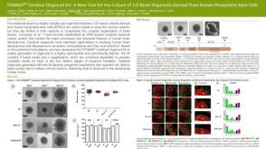 科学海报STEMdiff™ Cerebral Organoid Kit: A New Tool for the Culture of 3D Brain Organoids Derived from hPSCs

 沪公网安备31010102008431号