Pattison AM et al. (OCT 2016)
Infection and immunity 84 10 3083--91
Intestinal Enteroids Model Guanylate Cyclase C-Dependent Secretion Induced by Heat-Stable Enterotoxins.
Enterotoxigenic Escherichia coli (ETEC) causes 20% of the acute infectious diarrhea (AID) episodes worldwide,often by producing heat-stable enterotoxins (STs),which are peptides structurally homologous to paracrine hormones of the intestinal guanylate cyclase C (GUCY2C) receptor. While molecular mechanisms mediating ST-induced intestinal secretion have been defined,advancements in therapeutics have been hampered for decades by the paucity of disease models that integrate molecular and functional endpoints amenable to high-throughput screening. Here,we reveal that mouse and human intestinal enteroids in three-dimensional ex vivo cultures express the components of the GUCY2C secretory signaling axis. ST and its structural analog,linaclotide,an FDA-approved oral secretagog,induced fluid accumulation quantified simultaneously in scores of enteroid lumens,recapitulating ETEC-induced intestinal secretion. Enteroid secretion depended on canonical molecular signaling events responsible for ETEC-induced diarrhea,including cyclic GMP (cGMP) produced by GUCY2C,activation of cGMP-dependent protein kinase (PKG),and opening of the cystic fibrosis transmembrane conductance regulator (CFTR). Importantly,pharmacological inhibition of CFTR abrogated enteroid fluid secretion,providing proof of concept for the utility of this model to screen antidiarrheal agents. Intestinal enteroids offer a unique model,integrating the GUCY2C signaling axis and luminal fluid secretion,to explore the pathophysiology of,and develop platforms for,high-throughput drug screening to identify novel compounds to prevent and treat ETEC diarrheal disease.
View Publication
产品号#:
06005
产品名:
IntestiCult™ 类器官生长培养基 (小鼠)
Liu W et al. (OCT 2016)
Oncogene 35 40 5237--5247
Olfactomedin 4 deletion induces colon adenocarcinoma in Apc(Min/+) mice.
Colon carcinogenesis is a multiple-step process involving the accumulation of a series of genetic and epigenetic alterations. The most commonly initiating event of intestinal carcinogenesis is mutation of the adenomatous polyposis coli (APC) gene,which leads to activation of the Wnt/β-catenin pathway. Olfactomedin 4 (OLFM4) has emerged as an intestinal stem-cell marker,but its biological function in the intestine remains to be determined. Here we show that Olfm4 deletion induced colon adenocarcinoma in the distal colon of Apc(Min/+) mice. Mechanistically,we found that OLFM4 is a target gene of the Wnt/β-catenin pathway and can downregulate β-catenin signaling by competing with Wnt ligands for binding to Frizzled receptors,as well as by inhibition of the Akt-GSK-3β (Akt-glycogen synthase kinase-3β) pathway. We have shown that both Wnt and nuclear factor-κB (NF-κB) signaling were boosted in tumor tissues of Apc Olfm4 double-mutant mice. These data establish OLFM4 as a critical negative regulator of the Wnt/β-catenin and NF-κB pathways that inhibits colon-cancer development initiated by APC mutation. In addition,Olfm4 deletion significantly enhanced intestinal-crypt proliferation and inflammation induced by azoxymethane/dextran sodium sulfate. Thus,OLFM4 has an important role in the regulation of intestinal inflammation and tumorigenesis,and could be a potential therapeutic target for intestinal malignant tumors. Unlike the human colonic epithelium,the mouse colonic epithelium does not express OLFM4,but nevertheless,systemic OLFM4 deletion promotes colon tumorigenesis and that loss from mucosal neutrophils may have a role to play.
View Publication
产品号#:
06005
产品名:
IntestiCult™ 类器官生长培养基 (小鼠)
Alison MR et al. (DEC 2010)
The Journal of pathology 222 4 335--44
Finding cancer stem cells: are aldehyde dehydrogenases fit for purpose?
Despite many years of intensive effort,there is surprisingly little consensus on the most suitable markers with which to locate and isolate stem cells from adult tissues. By comparison,the study of cancer stem cells is still in its infancy; so,unsurprisingly,there is great uncertainty as to the identity of these cells. Stem cell markers can be broadly categorized into molecular determinants of self-renewal,clonogenicity,multipotentiality,adherence to the niche,and longevity. This review assesses the utility of recognizing cancer stem cells by virtue of high expression of aldehyde dehydrogenases (ALDHs),probably significant determinants of cell survival through their ability to detoxify many potentially cytotoxic molecules,and contributing to drug resistance. Antibodies are available against the ALDH enzyme family,but the vast majority of studies have used cell sorting techniques to enrich for cells expressing these enzymes. Live cells expressing high ALDH activity are usually identified by the ALDEFLUOR kit and sorted by fluorescence activated cell sorting (FACS). For many human tumours,but notably breast cancer,cell selection based upon ALDH activity appears to be a useful marker for enriching for cells with tumour-initiating activity (presumed cancer stem cells) in immunodeficient mice,and indeed the frequency of so-called ALDH(bri) cells in many tumours can be an independent prognostic indicator.
View Publication
产品号#:
01700
01705
01702
产品名:
ALDEFLUOR™ 试剂盒
ALDEFLUOR™ DEAB试剂, 1.5 mM, 1 mL
ALDEFLUOR™检测缓冲液
Taubert I et al. (APR 2011)
Cytotherapy 13 4 459--66
Characterization of hematopoietic stem cell subsets from patients with multiple myeloma after mobilization with plerixafor.
BACKGROUND AIMS: Previous studies have demonstrated that the combination of granulocyte-colony-stimulating factor (G-CSF) + plerixafor is more efficient in mobilizing CD34(+) hematopoietic stem cells (HSC) into the peripheral blood than G-CSF alone. In this study we analyzed the impact of adding plerixafor to G-CSF upon the mobilization of different HSC subsets. METHODS: We characterized the immunophenotype of HSC subsets isolated from the peripheral blood of eight patients with multiple myeloma (MM) before and after treatment with plerixafor. All patients were supposed to collect stem cells prior to high-dose chemotherapy and consecutive autologous stem cell transplantation,and therefore received front-line mobilization with 4 days of G-CSF followed by a single dose of plerixafor. Samples of peripheral blood were analyzed comparatively by flow cytometry directly before and 12 h after administration of plerixafor. RESULTS: The number of aldehyde dehydrogenase (ALDH)(bright) and CD34(+) cells was significantly higher after plerixafor treatment (1.2-5.0 and 1.5-6.0 times; both P textless 0.01) and an enrichment of the very primitive CD34(+) CD38(-) and ALDH(bright) CD34(+) CD38(-) HSC subsets was detectable. Additionally,two distinct ALDH(+) subsets could be clearly distinguished. The small ALDH(high) subset showed a higher number of CD34(+) CD38(-) cells in contrast to the total ALDH(bright) subpopulation and probably represented a very primitive subpopulation of HSC. CONCLUSIONS: A combined staining of ALDH,CD34 and CD38 might represent a powerful tool for the identification of a very rare and primitive hematopoietic stem cell subset. The addition of plerixafor mobilized not only more CD34(+) cells but was also able to increase the proportion of more primitive stem cell subsets.
View Publication
产品号#:
01700
01705
01702
产品名:
ALDEFLUOR™ 试剂盒
ALDEFLUOR™ DEAB试剂, 1.5 mM, 1 mL
ALDEFLUOR™检测缓冲液
Sullivan JP et al. (DEC 2010)
Cancer research 70 23 9937--48
Aldehyde dehydrogenase activity selects for lung adenocarcinoma stem cells dependent on notch signaling.
Aldehyde dehydrogenase (ALDH) is a candidate marker for lung cancer cells with stem cell-like properties. Immunohistochemical staining of a large panel of primary non-small cell lung cancer (NSCLC) samples for ALDH1A1,ALDH3A1,and CD133 revealed a significant correlation between ALDH1A1 (but not ALDH3A1 or CD133) expression and poor prognosis in patients including those with stage I and N0 disease. Flow cytometric analysis of a panel of lung cancer cell lines and patient tumors revealed that most NSCLCs contain a subpopulation of cells with elevated ALDH activity,and that this activity is associated with ALDH1A1 expression. Isolated ALDH(+) lung cancer cells were observed to be highly tumorigenic and clonogenic as well as capable of self-renewal compared with their ALDH(-) counterparts. Expression analysis of sorted cells revealed elevated Notch pathway transcript expression in ALDH(+) cells. Suppression of the Notch pathway by treatment with either a γ-secretase inhibitor or stable expression of shRNA against NOTCH3 resulted in a significant decrease in ALDH(+) lung cancer cells,commensurate with a reduction in tumor cell proliferation and clonogenicity. Taken together,these findings indicate that ALDH selects for a subpopulation of self-renewing NSCLC stem-like cells with increased tumorigenic potential,that NSCLCs harboring tumor cells with ALDH1A1 expression have inferior prognosis,and that ALDH1A1 and CD133 identify different tumor subpopulations. Therapeutic targeting of the Notch pathway reduces this ALDH(+) component,implicating Notch signaling in lung cancer stem cell maintenance.
View Publication
产品号#:
01700
01705
01702
产品名:
ALDEFLUOR™ 试剂盒
ALDEFLUOR™ DEAB试剂, 1.5 mM, 1 mL
ALDEFLUOR™检测缓冲液
Liu C et al. (MAY 2012)
Molecular biology reports 39 5 5875--81
Co-expression of Oct-4 and Nestin in human breast cancers.
The aim is to investigate the clinical implications of the Oct-4 and Nestin protein in human breast cancers. A total of 346 cases including 26 fresh and 320 paraffin-embedded tumor tissues were selected for characterizing the frequency of CD44(+)CD24(-) tumor cells by flow cytometry and the differential expression of the stem cell-related genes between CD44(+)CD24(-) and non-CD44(+)CD24(-) tumor cells was analyzed by PCR Array and immunofluorescence. In comparison with the non-CD44(+)CD24(-) tumor cells,the CD44(+)CD24(-),particularly for those with high percentage of Oct-4(+) and Nestin(+),tumor cells had higher tumorigenicity by forming mammospheres in vitro. More importantly,42 (13.125%) out of 320 tumor tissues were positive for Oct-4 and Nestin staining. Universal analysis and multivariate analysis revealed that the expression of Oct-4 and Nestin was associated significantly with younger age,pathogenic degrees,lymph node metastasis and triple-negative breast cancer independently (P textless 0.05) as well as shorter survival (P = 0.001). Oct-4 and Nestin were important regulators of the development of breast cancer,and Oct-4 and Nestin may be used as predictors for the prognosis of breast cancers.
View Publication
产品号#:
05620
产品名:
MammoCult™ 人源培养基套装
Zhu Y et al. (JAN 2013)
PLoS ONE 8 1 e54552
Three-Dimensional Neuroepithelial Culture from Human Embryonic Stem Cells and Its Use for Quantitative Conversion to Retinal Pigment Epithelium
A goal in human embryonic stem cell (hESC) research is the faithful differentiation to given cell types such as neural lineages. During embryonic development,a basement membrane surrounds the neural plate that forms a tight,apico-basolaterally polarized epithelium before closing to form a neural tube with a single lumen. Here we show that the three-dimensional epithelial cyst culture of hESCs in Matrigel combined with neural induction results in a quantitative conversion into neuroepithelial cysts containing a single lumen. Cells attain a defined neuroepithelial identity by 5 days. The neuroepithelial cysts naturally generate retinal epithelium,in part due to IGF-1/insulin signaling. We demonstrate the utility of this epithelial culture approach by achieving a quantitative production of retinal pigment epithelial (RPE) cells from hESCs within 30 days. Direct transplantation of this RPE into a rat model of retinal degeneration without any selection or expansion of the cells results in the formation of a donor-derived RPE monolayer that rescues photoreceptor cells. The cyst method for neuroepithelial differentiation of pluripotent stem cells is not only of importance for RPE generation but will also be relevant to the production of other neuronal cell types and for reconstituting complex patterning events from three-dimensional neuroepithelia.
View Publication
产品号#:
05854
05855
05850
05857
05870
05875
07913
85850
85857
85870
85875
产品名:
mFreSR™
mFreSR™
Dispase(5 U/mL)
mTeSR™1
mTeSR™1
Zhu S et al. (JUN 2017)
Nature 546 7660 667--670
Nlrp9b inflammasome restricts rotavirus infection in intestinal epithelial cells.
Rotavirus,a leading cause of severe gastroenteritis and diarrhoea in young children,accounts for around 215,000 deaths annually worldwide. Rotavirus specifically infects the intestinal epithelial cells in the host small intestine and has evolved strategies to antagonize interferon and NF-κB signalling,raising the question as to whether other host factors participate in antiviral responses in intestinal mucosa. The mechanism by which enteric viruses are sensed and restricted in vivo,especially by NOD-like receptor (NLR) inflammasomes,is largely unknown. Here we uncover and mechanistically characterize the NLR Nlrp9b that is specifically expressed in intestinal epithelial cells and restricts rotavirus infection. Our data show that,via RNA helicase Dhx9,Nlrp9b recognizes short double-stranded RNA stretches and forms inflammasome complexes with the adaptor proteins Asc and caspase-1 to promote the maturation of interleukin (Il)-18 and gasdermin D (Gsdmd)-induced pyroptosis. Conditional depletion of Nlrp9b or other inflammasome components in the intestine in vivo resulted in enhanced susceptibility of mice to rotavirus replication. Our study highlights an important innate immune signalling pathway that functions in intestinal epithelial cells and may present useful targets in the modulation of host defences against viral pathogens.
View Publication
产品号#:
06005
产品名:
IntestiCult™ 类器官生长培养基 (小鼠)
J. Wang et al. (JAN 2018)
Gastroenterology 154 6 1737--1750
Ceragenin CSA13 Reduces Clostridium difficile Infection in Mice by Modulating the Intestinal Microbiome and Metabolites.
BACKGROUND & AIMS Clostridium difficile induces intestinal inflammation by releasing toxins A and B. The antimicrobial compound cationic steroid antimicrobial 13 (CSA13) has been developed for treating gastrointestinal infections. The CSA13-Eudragit formulation can be given orally and releases CSA13 in the terminal ileum and colon. We investigated whether this form of CSA13 reduces C difficile infection (CDI) in mice. METHODS C57BL/6J mice were infected with C difficile on day 0,followed by subcutaneous administration of pure CSA13 or oral administration of CSA13-Eudragit (10 mg/kg/d for 10 days). Some mice were given intraperitoneal vancomycin (50 mg/kg daily) on days 0-4 and relapse was measured after antibiotic withdrawal. The mice were monitored until day 20; colon and fecal samples were collected on day 3 for analysis. Blood samples were collected for flow cytometry analyses. Fecal pellets were collected each day from mice injected with CSA13 and analyzed by high-performance liquid chromatography or 16S sequencing; feces were also homogenized in phosphate-buffered saline and fed to mice with CDI via gavage. RESULTS CDI of mice caused 60{\%} mortality,significant bodyweight loss,and colonic damage 3 days after infection; these events were prevented by subcutaneous injection of CSA13 or oral administration CSA13-Eudragit. There was reduced relapse of CDI after administration of CSA13 was stopped. Levels of CSA13 in feces from mice given CSA13-Eudragit were significantly higher than those of mice given subcutaneous CSA13. Subcutaneous and oral CSA13 each significantly increased the abundance of Peptostreptococcaceae bacteria and reduced the abundance of C difficile in fecal samples of mice. When feces from mice with CDI and given CSA13 were fed to mice with CDI that had not received CSA13,the recipient mice had significantly increased rates of survival. CSA13 reduced fecal levels of inflammatory metabolites (endocannabinoids) and increased fecal levels of 4 protective metabolites (ie,citrulline,3-aminoisobutyric acid,retinol,and ursodeoxycholic acid) in mice with CDI. Oral administration of these CSA13-dependent protective metabolites reduced the severity of CDI. CONCLUSIONS In studies of mice,we found the CSA13-Eudragit formulation to be effective in eradicating CDI by modulating the intestinal microbiota and metabolites.
View Publication

 EasySep™小鼠TIL(CD45)正选试剂盒


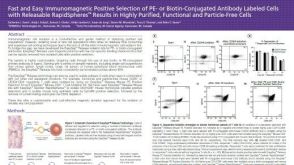 科学海报Positive Selection of PE- or Biotin-Conjugated Antibody Labeled Cells with Releasable Rapidspheres™

 沪公网安备31010102008431号