Brandl C et al. (SEP 2014)
NeuroMolecular Medicine 16 3 551--564
In-depth characterisation of Retinal Pigment Epithelium (RPE) cells derived from human induced pluripotent stem cells (hiPSC).
Induced pluripotent stem cell (iPSC)-derived retinal pigment epithelium (RPE) has widely been appreciated as a promising tool to model human ocular disease emanating from primary RPE pathology. Here,we describe the successful reprogramming of adult human dermal fibroblasts to iPSCs and their differentiation to pure expandable RPE cells with structural and functional features characteristic for native RPE. Fibroblast cultures were established from skin biopsy material and subsequently reprogrammed following polycistronic lentiviral transduction with OCT4,SOX2,KLF4 and L-Myc. Fibroblast-derived iPSCs showed typical morphology,chromosomal integrity and a distinctive stem cell marker profile. Subsequent differentiation resulted in expandable pigmented hexagonal RPE cells. The cells revealed stable RNA expression of mature RPE markers RPE65,RLBP and BEST1. Immunolabelling verified localisation of BEST1 at the basolateral plasma membrane,and scanning electron microscopy showed typical microvilli at the apical side of iPSC-derived RPE cells. Transepithelial resistance was maintained at high levels during cell culture indicating functional formation of tight junctions. Secretion capacity was demonstrated for VEGF-A. Feeding of porcine photoreceptor outer segments revealed the proper ability of these cells for phagocytosis. IPSC-derived RPE cells largely maintained these properties after cryopreservation. Together,our study underlines that adult dermal fibroblasts can serve as a valuable resource for iPSC-derived RPE with characteristics highly reminiscent of true RPE cells. This will allow its broad application to establish cellular models for RPE-related human diseases.
View Publication
产品号#:
05850
05857
05870
05875
07923
07930
07931
07940
07955
07956
07959
07954
85850
85857
85870
85875
100-1061
07952
产品名:
Dispase (1 U/mL)
CryoStor® CS10
CryoStor® CS10
CryoStor® CS10
CryoStor® CS10
CryoStor® CS10
mTeSR™1
mTeSR™1
CryoStor® CS10
CryoStor® CS10
Ji H et al. (JAN 2015)
The Journal of allergy and clinical immunology 135 1 236--244
Dynamic transcriptional and epigenomic reprogramming from pediatric nasal epithelial cells to induced pluripotent stem cells
BACKGROUND Induced pluripotent stem cells (iPSCs) hold tremendous potential,both as a biological tool to uncover the pathophysiology of disease by creating relevant human cell models and as a source of cells for cell-based therapeutic applications. Studying the reprogramming process will also provide significant insight into tissue development. OBJECTIVE We sought to characterize the derivation of iPSC lines from nasal epithelial cells (NECs) isolated from nasal mucosa samples of children,a highly relevant and easily accessible tissue for pediatric populations. METHODS We performed detailed comparative analysis on the transcriptomes and methylomes of NECs,iPSCs derived from NECs (NEC-iPSCs),and embryonic stem cells (ESCs). RESULTS NEC-iPSCs express pluripotent cell markers,can differentiate into all 3 germ layers in vivo and in vitro,and have a transcriptome and methylome remarkably similar to those of ESCs. However,residual DNA methylation marks exist,which are differentially methylated between NEC-iPSCs and ESCs. A subset of these methylation markers related to epithelium development and asthma and specific to NEC-iPSCs persisted after several passages in vitro,suggesting the retention of an epigenetic memory of their tissue of origin. Our analysis also identified novel candidate genes with dynamic gene expression and DNA methylation changes during reprogramming,which are indicative of possible roles in airway epithelium development. CONCLUSION NECs are an excellent tissue source to generate iPSCs in pediatric asthmatic patients,and detailed characterization of the resulting iPSC lines would help us better understand the reprogramming process and retention of epigenetic memory.
View Publication
产品号#:
05850
05857
05870
05875
85850
85857
85870
85875
产品名:
mTeSR™1
mTeSR™1
Wu H et al. (SEP 2011)
Journal of breast cancer 14 3 175--80
Can CD44+/CD24- Tumor Cells Be Used to Determine the Extent of Breast Cancer Invasion Following Neoadjuvant Chemotherapy?
PURPOSE: To investigate the distribution of CD44(+)/CD24(-) cells in breast cancers in relation to tumor size before and after the administration of neoadjuvant chemotherapy. METHODS: CD44(+)/CD24(-) tumor cells obtained from breast cancer specimens were characterized in vivo and in vitro using tumor formation assays and mammosphere generation assays,respectively. The distribution of CD44+/CD24- tumor cells in 78 breast cancer specimens following administration of neoadjuvant chemotherapy was also evaluated using immunofluorescence assays,and this distribution was compared with the extent of tumor invasion predicted by Response Evaluation Criteria in Solid Tumours (RECIST). RESULTS: In 27/78 cases,complete remission (CR) was identified using RECIST. However,18 of these CR cases were associated with a scattered distribution of tumor stem cells in the outline of the original tumor prior to neoadjuvant chemotherapy. After neoadjuvant chemotherapy,24 cases involved cancer cells that were confined to the tumor outline,and 21 cases had tumor cells or tumor stem cells overlapping the tumor outline. In addition,there were 6 patients who were insensitive to chemotherapy,and in these cases,both cancer cells and stem cells were detected outside the contours of the tumor volume imaged prior to chemotherapy. CONCLUSION: CD44+/CD24- tumor cells may be an additional parameter to evaluate when determining the extent of breast cancer invasion.
View Publication
产品号#:
05620
产品名:
MammoCult™ 人源培养基套装
Banerjee A et al. (JUL 2016)
Oncotarget 7 27 41432--41444
Endoplasmic reticulum stress and IRE-1 signaling cause apoptosis in colon cancer cells in response to andrographolide treatment
Bhushal S et al. ( 2017)
Frontiers in immunology 8 JUN 671
Cell Polarization and Epigenetic Status Shape the Heterogeneous Response to Type III Interferons in Intestinal Epithelial Cells.
Type I and type III interferons (IFNs) are crucial components of the first-line antiviral host response. While specific receptors for both IFN types exist,intracellular signaling shares the same Jak-STAT pathway. Due to its receptor expression,IFN-λ responsiveness is restricted mainly to epithelial cells. Here,we display IFN-stimulated gene induction at the single cell level to comparatively analyze the activities of both IFN types in intestinal epithelial cells and mini-gut organoids. Initially,we noticed that the response to both types of IFNs at low concentrations is based on a single cell decision-making determining the total cell intrinsic antiviral activity. We identified histone deacetylase (HDAC) activity as a crucial restriction factor controlling the cell frequency of IFN-stimulated gene (ISG) induction upon IFN-λ but not IFN-β stimulation. Consistently,HDAC blockade confers antiviral activity to an elsewise non-responding subpopulation. Second,in contrast to the type I IFN system,polarization of intestinal epithelial cells strongly enhances their ability to respond to IFN-λ signaling and raises the kinetics of gene induction. Finally,we show that ISG induction in mini-gut organoids by low amounts of IFN is characterized by a scattered heterogeneous responsiveness of the epithelial cells and HDAC activity fine-tunes exclusively IFN-λ activity. This study provides a comprehensive description of the differential response to type I and type III IFNs and demonstrates that cell polarization in gut epithelial cells specifically increases IFN-λ activity.
View Publication

 EasySep™小鼠TIL(CD45)正选试剂盒


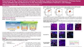 科学海报PneumaCult™-Ex Plus, a Novel Defined and Feeder-Free Medium, Supports the Improved Expansion of Primary Human Airway Epithelial Cells
 专家访谈Tamara Zietek, PhD Studying Intestinal Nutrient Absorption with Organoids
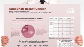 挂图SnapShot: Breast Cancer Overview of signaling pathways, commonly mutated genes and breast cancer subtypes

 沪公网安备31010102008431号