Y. Bhattarai et al. (JUN 2018)
Cell host & microbe 23 6 775--785.e5
Gut Microbiota-Produced Tryptamine Activates an Epithelial G-Protein-Coupled Receptor to Increase Colonic Secretion.
Tryptamine,a tryptophan-derived monoamine similar to 5-hydroxytryptamine (5-HT),is produced by gut bacteria and is abundant in human and rodent feces. However,the physiologic effect of tryptamine in the gastrointestinal (GI) tract remains unknown. Here,we show that the biological effects of tryptamine are mediated through the 5-HT4 receptor (5-HT4R),a G-protein-coupled receptor (GPCR) uniquely expressed in the colonic epithelium. Tryptamine increases both ionic flux across the colonic epithelium and fluid secretion in colonoids from germ-free (GF) and humanized (ex-GF colonized with human stool) mice,consistent with increased intestinal secretion. The secretory effect of tryptamine is dependent on 5-HT4R activation and is blocked by 5-HT4R antagonist and absent in 5-HT4R-/- mice. GF mice colonized by Bacteroides thetaiotaomicron engineered to produce tryptamine exhibit accelerated GI transit. Our study demonstrates an aspect of host physiology under control of a bacterial metabolite that can be exploited as a therapeutic modality. VIDEO ABSTRACT.
View Publication
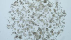
产品号#:
06005
产品名:
IntestiCult™ 类器官生长培养基 (小鼠)
M. D. Hu et al. (JUL 2018)
Journal of immunology (Baltimore,Md. : 1950) 201 2 747--756
Epithelial IL-15 Is a Critical Regulator of gamma$delta$ Intraepithelial Lymphocyte Motility within the Intestinal Mucosa.
Intraepithelial lymphocytes (IELs) expressing the gamma$delta$ TCR (gamma$delta$ IELs) provide continuous surveillance of the intestinal epithelium. However,the mechanisms regulating the basal motility of these cells within the epithelial compartment have not been well defined. We investigated whether IL-15 contributes to gamma$delta$ IEL localization and migratory behavior in addition to its role in IEL differentiation and survival. Using advanced live cell imaging techniques in mice,we find that compartmentalized overexpression of IL-15 in the lamina propria shifts the distribution of gamma$delta$ T cells from the epithelial compartment to the lamina propria. This mislocalization could be rescued by epithelial IL-15 overexpression,indicating that epithelial IL-15 is essential for gamma$delta$ IEL migration into the epithelium. Furthermore,in vitro analyses demonstrated that exogenous IL-15 stimulates gamma$delta$ IEL migration into cultured epithelial monolayers,and inhibition of IL-2Rbeta$ significantly attenuates the basal motility of these cells. Intravital microscopy showed that impaired IL-2Rbeta$ signaling induced gamma$delta$ IEL idling within the lateral intercellular space,which resulted in increased early pathogen invasion. Similarly,the redistribution of gamma$delta$ T cells to the lamina propria due to local IL-15 overproduction also enhanced bacterial translocation. These findings thus reveal a novel role for IL-15 in mediating gamma$delta$ T cell localization within the intestinal mucosa and regulating gamma$delta$ IEL motility and patrolling behavior as a critical component of host defense.
View Publication
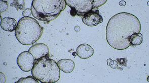
产品号#:
06005
产品名:
IntestiCult™ 类器官生长培养基 (小鼠)
C. L. Kraft et al. (NOV 2017)
Oncotarget 8 61 102923--102933
GUCY2C maintains intestinal LGR5+stem cells by opposing ER stress.
Long-lived multipotent stem cells (ISCs) at the base of intestinal crypts adjust their phenotypes to accommodate normal maintenance and post-injury regeneration of the epithelium. Their long life,lineage plasticity,and proliferative potential underlie the necessity for tight homeostatic regulation of the ISC compartment. In that context,the guanylate cyclase C (GUCY2C) receptor and its paracrine ligands regulate intestinal epithelial homeostasis,including proliferation,lineage commitment,and DNA damage repair. However,a role for this axis in maintaining ISCs remains unknown. Transgenic mice enabling analysis of ISCs (Lgr5-GFP) in the context of GUCY2C elimination (Gucy2c -/- ) were combined with immunodetection techniques and pharmacological treatments to define the role of the GUCY2C signaling axis in supporting ISCs. ISCs were reduced inGucy2c -/- mice,associated with loss of active Lgr5+cells but a reciprocal increase in reserve Bmi1+cells. GUCY2C was expressed in crypt base Lgr5+cells in which it mediates canonical cyclic (c) GMP-dependent signaling. Endoplasmic reticulum (ER) stress,typically absent from ISCs,was elevated throughout the crypt base inGucy2c -/- mice. The chemical chaperone tauroursodeoxycholic acid resolved this ER stress and restored the balance of ISCs,an effect mimicked by the GUCY2C effector 8Br-cGMP. Reduced ISCs inGucy2c -/- mice was associated with greater epithelial injury and impaired regeneration following sub-lethal doses of irradiation. These observations suggest that GUCY2C provides homeostatic signals that modulate ER stress and cell vulnerability as part of the machinery contributing to the integrity of ISCs.
View Publication
产品号#:
06005
产品名:
IntestiCult™ 类器官生长培养基 (小鼠)
B. Wang et al. (FEB 2018)
Cell stem cell 22 2 206--220.e4
Phospholipid Remodeling and Cholesterol Availability Regulate Intestinal Stemness and Tumorigenesis.
Adequate availability of cellular building blocks,including lipids,is a prerequisite for cellular proliferation,but excess dietary lipids are linked to increased cancer risk. Despite these connections,specific regulatory relationships between membrane composition,intestinal stem cell (ISC) proliferation,and tumorigenesis are unclear. We reveal an unexpected link between membrane phospholipid remodeling and cholesterol biosynthesis and demonstrate that cholesterol itself acts as a mitogen for ISCs. Inhibition of the phospholipid-remodeling enzyme Lpcat3 increases membrane saturation and stimulates cholesterol biosynthesis,thereby driving ISC proliferation. Pharmacologic inhibition of cholesterol synthesis normalizes crypt hyperproliferation in Lpcat3-deficient organoids and mice. Conversely,increasing cellular cholesterol content stimulates crypt organoid growth,and providing excess dietary cholesterol or driving endogenous cholesterol synthesis through SREBP-2 expression promotes ISC proliferation in vivo. Finally,disruption of Lpcat3-dependent phospholipid and cholesterol homeostasis dramatically enhances tumor formation in Apcminmice. These findings identify a critical dietary-responsive phospholipid-cholesterol axis regulating ISC proliferation and tumorigenesis.
View Publication
产品号#:
06005
产品名:
IntestiCult™ 类器官生长培养基 (小鼠)
Lungova V et al. ( 2014)
1307 237--243
Derivation of Epithelial Cells from Human Embryonic Stem Cells as an In Vitro Model of Vocal Mucosa
Vocal fold epithelial cells are very difficult to study as the vocal fold epithelial cell lines do not exist and they cannot be removed from the healthy larynx without engendering a significant and unacceptable risk to vocal fold function. Here,we describe the procedure to create an engineered vocal fold tissue construct consisting of the scaffold composed of the collagen 1 gel seeded with human fibroblasts and simple epithelial progenitors seeded on the scaffold and cultivated at air-liquid interface for 19-21 days to derive the stratified squamous epithelium. This model of vocal fold mucosa is very similar in morphology,gene expression,and phenotypic characteristics to native vocal fold epithelial cells and the underlying lamina propria and,therefore,offers a promising approach to studying vocal fold biology and biomechanics in health and disease.
View Publication
产品号#:
05850
05857
05870
05875
85850
85857
85870
85875
产品名:
mTeSR™1
mTeSR™1
Ling SSM et al. (JUN 2015)
PLOS ONE 10 6 e0131460
Instrumental Role of Helicobacter pylori γ-Glutamyl Transpeptidase in VacA-Dependent Vacuolation in Gastric Epithelial Cells
Helicobacter pylori causes cellular vacuolation in host cells,a cytotoxic event attributed to vacuolating cytotoxin (VacA) and the presence of permeant weak bases such as ammonia. We report here the role of γ-glutamyl transpeptidase (GGT),a constitutively expressed secretory enzyme of H. pylori,in potentiating VacA-dependent vacuolation formation in H. pylori-infected AGS and primary gastric cells. The enhancement is brought about by GGT hydrolysing glutamine present in the extracellular medium,thereby releasing ammonia which accentuates the VacA-induced vacuolation. The events of vacuolation in H. pylori wild type (WT)- and Δggt-infected AGS cells were first captured and visualized by real-time phase-contrast microscopy where WT was observed to induce more vacuoles than Δggt. By using semi-quantitative neutral red uptake assay,we next showed that Δggt induced significantly less vacuolation in AGS and primary gastric epithelial cells as compared to the parental strain (Ptextless0.05) indicating that GGT potentiates the vacuolating effect of VacA. Notably,vacuolation induced by WT was significantly reduced in the absence of GGT substrate,glutamine (Ptextless0.05) or in the presence of a competitive GGT inhibitor,serine-borate complex. Furthermore,the vacuolating ability of Δggt was markedly restored when co-incubated with purified recombinant GGT (rGGT),although rGGT itself did not induce vacuolation independently. Similarly,the addition of exogenous ammonium chloride as a source of ammonia also rescued the ability of Δggt to induce vacuolation. Additionally,we also show that monoclonal antibodies against GGT effectively inhibited GGT activity and successfully suppressed H. pylori-induced vacuolation. Collectively,our results clearly demonstrate that generation of ammonia by GGT through glutamine hydrolysis is responsible for enhancing VacA-dependent vacuolation. Our findings provide a new perspective on GGT as an important virulence factor and a promising target in the management of H. pylori-associated gastric diseases.
View Publication

 EasySep™小鼠TIL(CD45)正选试剂盒


 实验方案Cryopreserving Hepatic Organoids Expanded in HepatiCult™ Organoid Growth Medium (Human)
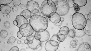 实验方案Recovery and Expansion of Hepatic Organoids Using HepatiCult™ Organoid Growth Medium
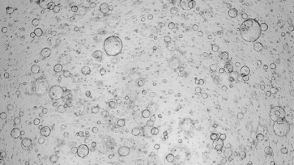 实验方案Expansion of Hepatic Organoids via Single Cells Using HepatiCult™ Organoid Growth Medium

 沪公网安备31010102008431号