Deonarain R et al. (NOV 2003)
Proceedings of the National Academy of Sciences of the United States of America 100 23 13453--8
Critical roles for IFN-beta in lymphoid development, myelopoiesis, and tumor development: links to tumor necrosis factor alpha.
We have generated mice null for IFN-beta and report the diverse consequences of IFN-beta for both the innate and adaptive arms of immunity. Despite no abnormalities in the proportional balance of CD4 and CD8 T cell populations in the peripheral blood,thymus,and spleen of IFN-beta-/- mice,activated lymph node and splenic T lymphocytes exhibit enhanced T cell proliferation and decreased tumor necrosis factor alpha production,relative to IFN-beta+/+ mice. Notably,constitutive and induced expression of tumor necrosis factor alpha is reduced in the spleen and bone marrow (BM) macrophages,respectively,of IFN-beta-/- mice. We also observe an altered splenic architecture in IFN-beta-/- mice and a reduction in resident macrophages. We identify a potential defect in B cell maturation in IFN-beta-/- mice,associated with a decrease in B220+ve/high/CD43-ve BM-derived cells and a reduction in BP-1,IgM,and CD23 expression. Circulating IgM-,Mac-1-,and Gr-1-positive cells are also substantially decreased in IFN-beta-/- mice. The decrease in the numbers of circulating macrophages and granulocytes likely reflects defective maturation of primitive BM hematopoiesis in mice,shown by the reduction of colony-forming units,granulocyte-macrophage. We proceeded to evaluate the in vivo growth of malignant cells in the IFN-beta-/- background and give evidence that Lewis lung carcinoma-specific tumor growth is more aggressive in IFN-beta-/- mice. Taken altogether,our data suggest that,in addition to the direct growth-inhibitory effects on tumor cells,IFN-beta is required during different stages of maturation in the development of the immune system.
View Publication
产品号#:
03434
03444
产品名:
MethoCult™ GF M3434
MethoCult™ GF M3434
Elling C et al. (MAR 2011)
Blood 117 10 2935--43
Novel imatinib-sensitive PDGFRA-activating point mutations in hypereosinophilic syndrome induce growth factor independence and leukemia-like disease.
The FIP1L1-PDGFRA fusion is seen in a fraction of cases with a presumptive diagnosis of hypereosinophilic syndrome (HES). However,because most HES patients lack FIP1L1-PDGFRA,we studied whether they harbor activating mutations of the PDGFRA gene. Sequencing of 87 FIP1L1-PDGFRA-negative HES patients revealed several novel PDGFRA point mutations (R481G,L507P,I562M,H570R,H650Q,N659S,L705P,R748G,and Y849S). When cloned into 32D cells,N659S and Y849S and-on selection for high expressors-also H650Q and R748G mutants induced growth factor-independent proliferation,clonogenic growth,and constitutive phosphorylation of PDGFRA and Stat5. Imatinib antagonized Stat5 phosphorylation. Mutations involving positions 659 and 849 had been shown previously to possess transforming potential in gastrointestinal stromal tumors. Because H650Q and R748G mutants possessed only weak transforming activity,we injected 32D cells harboring these mutants or FIP1L1-PDGFRA into mice and found that they induced a leukemia-like disease. Oral imatinib treatment significantly decreased leukemic growth in vivo and prolonged survival. In conclusion,our data provide evidence that imatinib-sensitive PDGFRA point mutations play an important role in the pathogenesis of HES and we propose that more research should be performed to further define the frequency and treatment response of PDGFRA mutations in FIP1L1-PDGFRA-negative HES patients.
View Publication
Schreiber A et al. (JUL 2005)
Journal of the American Society of Nephrology : JASN 16 7 2216--24
Membrane proteinase 3 expression in patients with Wegener's granulomatosis and in human hematopoietic stem cell-derived neutrophils.
A large membrane proteinase 3 (mPR3)-positive neutrophil subset (mPR3high) is a risk for Wegener's granulomatosis (WG). The relationship between mPR3 expression and clinical manifestations was investigated in 81 WG patients and mPR3 expression was studied in CD34+ stem cell-derived human neutrophils. The mPR3high neutrophil percentage correlated with renal function,anemia,and albumin at the time of presentation. The mPR3high neutrophil percentage and renal failure severity correlated directly after 5 yr. For elucidating mechanisms that govern mPR3 expression,studies were conducted to determine whether the genetic information that governs mPR3 expression resides within the neutrophils,even without stimuli possibly related to disease. CD34+ hematopoietic stem cells were differentiated to neutrophils,and their mPR3 expression was determined. A two-step amplification/differentiation protocol was used to differentiate human CD34+ hematopoietic stem cells into neutrophils with G-CSF. The cells progressively expressed the neutrophil surface markers CD66b,CD35,and CD11b. The ferricytochrome C assay demonstrated a strong respiratory burst at day 14 in response to PMA but none at day 0. Intracellular PR3 was detectable from day 4 by Western blotting. An increasing percentage of a mPR3-positive neutrophil subset became detectable by flow cytometry,whereas a second subset remained negative,consistent with a bimodal expression. Finally,human PR3-anti-neutrophil cytoplasmic autoantibodies induced a stronger respiratory burst,compared with human control IgG in stem cell-derived neutrophils. Taken together,these studies underscore the clinical importance of the WG mPR3 phenotype. The surface mPR3 on resting cells is probably genetically determined rather than being dictated by external factors.
View Publication
产品号#:
09600
09650
产品名:
StemSpan™ SFEM
StemSpan™ SFEM
Gibbs BF et al. (MAR 2008)
Clinical and experimental allergy : journal of the British Society for Allergy and Clinical Immunology 38 3 480--5
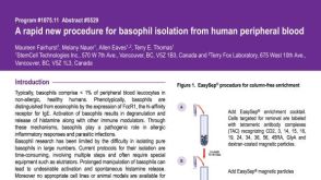
A rapid two-step procedure for the purification of human peripheral blood basophils to near homogeneity.
BACKGROUND: Basophils are increasingly utilized as indicators of allergic inflammation and as primary allergic effector cells to study signalling pathways. However,until the present,their enrichment has been time consuming,costly and limited to relatively few specialized laboratories. OBJECTIVE: We have therefore devised a reproducible and rapid method for the purification of human basophils from small quantities of peripheral blood within 1.5 h,which does not require the use of specialized equipment such as elutriators. METHODS: Human basophils were obtained from healthy volunteers undergoing venipuncture. Heparinized or K3-ethylenediaminetetraacetic acid blood samples were first subjected to centrifugation in Hetasep,directly followed by negative selection using immunomagnetic beads. Basophil morphology and purity were assessed by May-Grünwald staining of cytospins. IgE-mediated histamine release was analysed spectrofluorometrically and IL-4 and IL-13 production by quantitative RT-PCR. CD203c and CD63 surface expression was measured using flow cytometry before and after activation with anti-IgE. RESULTS: Using this protocol,basophils were enriched close to homogeneity in most cases with a mean purity of 99.34+/-0.88% (range 97-100%,n=18) and a mean recovery of 75.6 (range 39-100%,n=8). Basophil viability following purification was 99.6+/-0.89% using Trypan blue exclusion. The purification procedure gave rise to basophils with normal functional responses to anti-IgE regarding histamine release as well as IL-4 and IL-13 mRNA expression. Moreover,constitutive cell-surface CD203c/CD63 expressions were not elevated before anti-IgE stimulation. CONCLUSION: The rapidity,simplicity and reproducibility of this method will facilitate the employment of basophils in high-output ex vivo studies.
View Publication

 EasySep™小鼠TIL(CD45)正选试剂盒


 挂图Frequencies of Immune Cells in Rat Tissue Lists the estimated frequencies of more than 15 immune cell types in Sprague Dawley rats


 沪公网安备31010102008431号