Neves H et al. (MAY 2006)
Stem cells (Dayton,Ohio) 24 5 1328--37
Effects of Delta1 and Jagged1 on early human hematopoiesis: correlation with expression of notch signaling-related genes in CD34+ cells.
It has been shown that Notch signaling mediated by ligands of both Jagged and Delta families expands the hematopoietic stem cell compartment while blocking or delaying terminal myeloid differentiation. Here we show that Delta1- and Jagged1-expressing stromal cells have distinct effects on the clonogenic and differentiation capacities of human CD34(+) CD38(+) cells. Jagged1 increases the number of bipotent colony-forming unit-granulocyte macrophage (CFU-GM) and unipotent progenitors (CFU-granulocytes and CFU-macrophages),without quantitatively affecting terminal cell differentiation,whereas Delta1 reduces the number of CFU-GM and differentiated monocytic cells. Expression analysis of genes coding for Notch receptors,Notch targets,and Notch signaling modulators in supernatant CD34(+) cells arising upon contact with Jagged1 and Delta1 shows dynamic and differential gene expression profiles over time. At early time points,modest upregulation of Notch1,Notch3,and Hes1 was observed in Jagged1-CD34(+) cells,whereas those in contact with Delta1 strikingly upregulated Notch3 and Hes1. Later,myeloid progenitors with strong clonogenic potential emerging upon contact with Jagged1 upregulated Notch1 and Deltex and downregulated Notch signaling modulators,whereas T/NK progenitors originated by Delta1 strikingly upregulated Notch3 and Deltex and,to a lesser extent,Hes1,Lunatic Fringe,and Numb. Together,the data unravel previously unrecognized expression patterns of Notch signaling-related genes in CD34(+) CD38(+) cells as they develop in Jagged1- or Delta1-stromal cell environments,which appear to reflect sequential maturational stages of CD34(+) cells into distinct cell lineages.
View Publication
产品号#:
04435
04445
产品名:
MethoCult™ H4435 Enriched
MethoCult™ H4435 Enriched
Senn JJ et al. (SEP 2005)
The Journal of pharmacology and experimental therapeutics 314 3 972--9
Non-CpG-containing antisense 2'-methoxyethyl oligonucleotides activate a proinflammatory response independent of Toll-like receptor 9 or myeloid differentiation factor 88.
Oligonucleotides with a CpG" motif trigger a proinflammatory response through activation of Toll-like receptor 9 (TLR9) and are being studied to exploit these properties for use as adjuvants and cancer therapies. However�
View Publication
产品号#:
09600
09650
产品名:
StemSpan™ SFEM
StemSpan™ SFEM
Karp JE et al. (MAY 2009)
Blood 113 20 4841--52
Active oral regimen for elderly adults with newly diagnosed acute myelogenous leukemia: a preclinical and phase 1 trial of the farnesyltransferase inhibitor tipifarnib (R115777, Zarnestra) combined with etoposide.
The farnesyltransferase inhibitor tipifarnib exhibits modest activity against acute myelogenous leukemia. To build on these results,we examined the effect of combining tipifarnib with other agents. Tipifarnib inhibited signaling downstream of the farnesylated small G protein Rheb and synergistically enhanced etoposide-induced antiproliferative effects in lymphohematopoietic cell lines and acute myelogenous leukemia isolates. We subsequently conducted a phase 1 trial of tipifarnib plus etoposide in adults over 70 years of age who were not candidates for conventional therapy. A total of 84 patients (median age,77 years) received 224 cycles of oral tipifarnib (300-600 mg twice daily for 14 or 21 days) plus oral etoposide (100-200 mg daily on days 1-3 and 8-10). Dose-limiting toxicities occurred with 21-day tipifarnib. Complete remissions were achieved in 16 of 54 (30%) receiving 14-day tipifarnib versus 5 of 30 (17%) receiving 21-day tipifarnib. Complete remissions occurred in 50% of two 14-day tipifarnib cohorts: 3A (tipifarnib 600,etoposide 100) and 8A (tipifarnib 400,etoposide 200). In vivo,tipifarnib plus etoposide decreased ribosomal S6 protein phosphorylation and increased histone H2AX phosphorylation and apoptosis. Tipifarnib plus etoposide is a promising orally bioavailable regimen that warrants further evaluation in elderly adults who are not candidates for conventional induction chemotherapy. These clinical studies are registered at www.clinicaltrials.gov as NCT00112853.
View Publication
产品号#:
04434
04444
产品名:
MethoCult™ H4434 Classic
MethoCult™ H4434 Classic
Baba Y et al. (AUG 2006)
Journal of immunology (Baltimore,Md. : 1950) 177 4 2294--303
Constitutively active beta-catenin promotes expansion of multipotent hematopoietic progenitors in culture.
This study was designed to investigate one component of the Wnt/beta-catenin signaling pathway that has been implicated in stem cell self-renewal. Retroviral-mediated introduction of stable beta-catenin to primitive murine bone marrow cells allowed the expansion of multipotential c-Kit(low)Sca-1(low/-)CD19(-) CD11b/Mac-1(-)Flk-2(-)CD43(+)AA4.1(+)NK1.1(-)CD3(-)CD11c(-)Gr-1(-)CD45R/B220(+) cells in the presence of stromal cells and cytokines. They generated myeloid,T,and B lineage lymphoid cells in culture,but had no T lymphopoietic potential when transplanted. Stem cell factor and IL-6 were found to be minimal requirements for long-term,stromal-free propagation,and a beta-catenin-transduced cell line was maintained for 5 mo with these defined conditions. Although multipotential and responsive to many normal stimuli in culture,it was unable to engraft several types of irradiated recipients. These findings support previous studies that have implicated the canonical Wnt pathway signaling in regulation of multipotent progenitors. In addition,we demonstrate how it may be experimentally manipulated to generate valuable cell lines.
View Publication
产品号#:
03434
03444
产品名:
MethoCult™ GF M3434
MethoCult™ GF M3434
Zhao Z et al. (JUL 2010)
Genes & development 24 13 1389--402
p53 loss promotes acute myeloid leukemia by enabling aberrant self-renewal.
The p53 tumor suppressor limits proliferation in response to cellular stress through several mechanisms. Here,we test whether the recently described ability of p53 to limit stem cell self-renewal suppresses tumorigenesis in acute myeloid leukemia (AML),an aggressive cancer in which p53 mutations are associated with drug resistance and adverse outcome. Our approach combined mosaic mouse models,Cre-lox technology,and in vivo RNAi to disable p53 and simultaneously activate endogenous Kras(G12D)-a common AML lesion that promotes proliferation but not self-renewal. We show that p53 inactivation strongly cooperates with oncogenic Kras(G12D) to induce aggressive AML,while both lesions on their own induce T-cell malignancies with long latency. This synergy is based on a pivotal role of p53 in limiting aberrant self-renewal of myeloid progenitor cells,such that loss of p53 counters the deleterious effects of oncogenic Kras on these cells and enables them to self-renew indefinitely. Consequently,myeloid progenitor cells expressing oncogenic Kras and lacking p53 become leukemia-initiating cells,resembling cancer stem cells capable of maintaining AML in vivo. Our results establish an efficient new strategy for interrogating oncogene cooperation,and provide strong evidence that the ability of p53 to limit aberrant self-renewal contributes to its tumor suppressor activity.
View Publication
产品号#:
03534
09600
09650
产品名:
MethoCult™ GF M3534
StemSpan™ SFEM
StemSpan™ SFEM
Maes C et al. (MAY 2006)
The Journal of clinical investigation 116 5 1230--42
Placental growth factor mediates mesenchymal cell development, cartilage turnover, and bone remodeling during fracture repair.
Current therapies for delayed- or nonunion bone fractures are still largely ineffective. Previous studies indicated that the VEGF homolog placental growth factor (PlGF) has a more significant role in disease than in health. Therefore we investigated the role of PlGF in a model of semi-stabilized bone fracture healing. Fracture repair in mice lacking PlGF was impaired and characterized by a massive accumulation of cartilage in the callus,reminiscent of delayed- or nonunion fractures. PlGF was required for the early recruitment of inflammatory cells and the vascularization of the fracture wound. Interestingly,however,PlGF also played a role in the subsequent stages of the repair process. Indeed in vivo and in vitro findings indicated that PlGF induced the proliferation and osteogenic differentiation of mesenchymal progenitors and stimulated cartilage turnover by particular MMPs. Later in the process,PlGF was required for the remodeling of the newly formed bone by stimulating osteoclast differentiation. As PlGF expression was increased throughout the process of bone repair and all the important cell types involved expressed its receptor VEGFR-1,the present data suggest that PlGF is required for mediating and coordinating the key aspects of fracture repair. Therefore PlGF may potentially offer therapeutic advantages for fracture repair.
View Publication


 EasySep™小鼠TIL(CD45)正选试剂盒




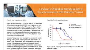 产品手册Services for Predicting Hematotoxicity in Drug Development with HemaTox™ Assays




 沪公网安备31010102008431号