Stutz MD et al. (DEC 2017)
Cell death and differentiation
Necroptotic signaling is primed in Mycobacterium tuberculosis-infected macrophages, but its pathophysiological consequence in disease is restricted.
Mixed lineage kinase domain-like (MLKL)-dependent necroptosis is thought to be implicated in the death of mycobacteria-infected macrophages,reportedly allowing escape and dissemination of the microorganism. Given the consequent interest in developing inhibitors of necroptosis to treat Mycobacterium tuberculosis (Mtb) infection,we used human pharmacologic and murine genetic models to definitively establish the pathophysiological role of necroptosis in Mtb infection. We observed that Mtb infection of macrophages remodeled the intracellular signaling landscape by upregulating MLKL,TNFR1,and ZBP1,whilst downregulating cIAP1,thereby establishing a strong pro-necroptotic milieu. However,blocking necroptosis either by deleting Mlkl or inhibiting RIPK1 had no effect on the survival of infected human or murine macrophages. Consistent with this,MLKL-deficiency or treatment of humanized mice with the RIPK1 inhibitor Nec-1s did not impact on disease outcomes in vivo,with mice displaying lung histopathology and bacterial burdens indistinguishable from controls. Therefore,although the necroptotic pathway is primed by Mtb infection,macrophage necroptosis is ultimately restricted to mitigate disease pathogenesis. We identified cFLIP upregulation that may promote caspase 8-mediated degradation of CYLD,and other necrosome components,as a possible mechanism abrogating Mtb's capacity to coopt necroptotic signaling. Variability in the capacity of these mechanisms to interfere with necroptosis may influence disease severity and could explain the heterogeneity of Mtb infection and disease.
View Publication
Brandl M et al. (AUG 1999)
Experimental hematology 27 8 1264--70
Bispecific antibody fragments with CD20 X CD28 specificity allow effective autologous and allogeneic T-cell activation against malignant cells in peripheral blood and bone marrow cultures from patients with B-cell lineage leukemia and lymphoma.
Bispecific antibodies directed against tumor-associated target antigens and to surface receptors mediating T-cell activation,such as the TCR/CD3 complex and the costimulatory receptor CD28,are capable of mediating T-cell activation resulting in tumor cell killing. In this study,we used the B-cell-associated antigens CD19 and CD20 as target structures on human leukemic cells. We found that a combination of bispecific antibody fragments (bsFab2) with target x CD3 and target x CD28 specificity induces vigorous autologous T-cell activation and killing of malignant cells in peripheral blood and bone marrow cultures from patients with chronic lymphocytic leukemia and follicular lymphoma. The bsFab2 targeting CD20 were considerably more effective than those binding to CD19. The colony-forming capacity of treated bone marrow was impaired due to large amounts of tumor necrosis factor alpha produced during bsFab2-induced T-cell activation. Neutralizing tumor necrosis factor alpha antibodies were found to reverse this negative effect without affecting T-cell activation and tumor cell killing. CD20 x CD28 bsFab2,when used alone rather than in combination,markedly improved the recognition of leukemic cells by allogeneic T cells. Therefore,these reagents may be capable of enhancing the immunogenicity of leukemic cells in general and,in particular,of increasing the antileukemic activity of allogeneic donor buffy coat cells in relapsed bone marrow transplanted patients.
View Publication
产品号#:
04431
产品名:
MethoCult™ H4431
Osada H et al. (APR 2001)
Transfusion 41 4 499--503
Detection of fetal HPCs in maternal circulation after delivery.
BACKGROUND: Circulation of mature fetal blood cells in the maternal blood for a certain postpartum period has been verified,but detailed study of the fetal HPCs has not been reported. The objective of this study was to evaluate the frequency and clearance of these cells in the peripheral blood of puerperal women. STUDY DESIGN AND METHODS: PBMNCs from 15 puerperal women who gave birth to male infants were cultured in semi-solid medium containing hematopoietic stimulating factors. Colonies formed in the medium were individually characterized,collected,and subjected to PCR amplification of the SRY gene on Y chromosome to confirm fetal origin. RESULTS: The mean numbers of fetal progenitor cell colonies isolated per mL of maternal blood were 1.63,2.48,0.56,0.12,and 0 on the day of delivery,at 4 days,1 month,6 months,and 1 year after delivery,respectively. There was no difference in the ratio of fetal versus maternal colonies between erythroid and granulocyte/macrophage lineages. CONCLUSION: The present study demonstrated that a significant number of fetal HPCs circulate in the maternal blood for a duration of at least 6 months after delivery.
View Publication

 EasySep™小鼠TIL(CD45)正选试剂盒


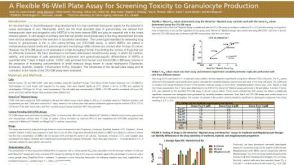 科学海报A Flexible 96-Well Plate Assay for Screening Toxicity to Granulocyte Production
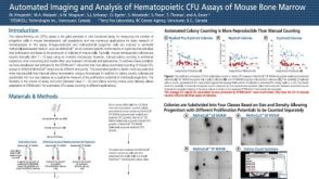 科学海报Automated Imaging and Analysis of Hematopoietic CFU Assays of Mouse Bone Marrow
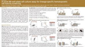 科学海报A Novel 96-well Plate Cell Culture Assay for Lineage-Specific Hematopoietic Cell Toxicity Screening


 沪公网安备31010102008431号