ADA-deficient SCID is associated with a specific microenvironment and bone phenotype characterized by RANKL/OPG imbalance and osteoblast insufficiency.
Adenosine deaminase (ADA) deficiency is a disorder of the purine metabolism leading to combined immunodeficiency and systemic alterations,including skeletal abnormalities. We report that ADA deficiency in mice causes a specific bone phenotype characterized by alterations of structural properties and impaired mechanical competence. These alterations are the combined result of an imbalanced receptor activator of nuclear factor-kappaB ligand (RANKL)/osteoprotegerin axis,causing decreased osteoclastogenesis and an intrinsic defect of osteoblast function with subsequent low bone formation. In vitro,osteoblasts lacking ADA displayed an altered transcriptional profile and growth reduction. Furthermore,the bone marrow microenvironment of ADA-deficient mice showed a reduced capacity to support in vitro and in vivo hematopoiesis. Treatment of ADA-deficient neonatal mice with enzyme replacement therapy,bone marrow transplantation,or gene therapy resulted in full recovery of the altered bone parameters. Remarkably,untreated ADA-severe combined immunodeficiency patients showed a similar imbalance in RANKL/osteoprotegerin levels alongside severe growth retardation. Gene therapy with ADA-transduced hematopoietic stem cells increased serum RANKL levels and children's growth. Our results indicate that the ADA metabolism represents a crucial modulatory factor of bone cell activities and remodeling.
View Publication
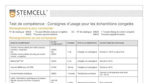
产品号#:
13056
产品名:
Iversen PO et al. (MAR 2010)
American journal of physiology. Regulatory,integrative and comparative physiology 298 3 R808--14
Separate mechanisms cause anemia in ischemic vs. nonischemic murine heart failure.
In ischemic congestive heart failure (CHF),anemia is associated with poor prognosis. Whether anemia develops in nonischemic CHF is uncertain. The hematopoietic inhibitors TNF-alpha and nitric oxide (NO) are activated in ischemic CHF. We examined whether mice with ischemic or nonischemic CHF develop anemia and whether TNF-alpha and NO are involved. We studied mice (n = 7-9 per group) with CHF either due to myocardial infarction (MI) or to overexpression of the Ca(2+)-binding protein calsequestrin (CSQ) or to induced cardiac disruption of the sarcoplasmic reticulum Ca(2+)-ATPase 2 gene (SERCA2 KO). Hematopoiesis was analyzed by colony formation of CD34(+) bone marrow cells. Hemoglobin concentration was 14.0 +/- 0.4 g/dl (mean +/- SD) in controls,while it was decreased to 10.1 +/- 0.4,9.7 +/- 0.4,and 9.6 +/- 0.3 g/dl in MI,CSQ,and SERCA2 KO,respectively (P textless 0.05). Colony numbers per 100,000 CD34(+) cells in the three CHF groups were reduced to 33 +/- 3 (MI),34 +/- 3 (CSQ),and 39 +/- 3 (SERCA2 KO) compared with 68 +/- 4 in controls (P textless 0.05). Plasma TNF-alpha nearly doubled in MI,and addition of anti-TNF-alpha antibody normalized colony formation. Inhibition of colony formation was completely abolished with blockade of endothelial NO synthase in CSQ and SERCA2 KO,but not in MI. In conclusion,the mechanism of anemia in CHF depends on the etiology of cardiac disease; whereas TNF-alpha impairs hematopoiesis in CHF following MI,NO inhibits blood cell formation in nonischemic murine CHF.
View Publication
产品号#:
产品名:
Randrianarison-Huetz V et al. (APR 2010)
Blood 115 14 2784--95
Gfi-1B controls human erythroid and megakaryocytic differentiation by regulating TGF-beta signaling at the bipotent erythro-megakaryocytic progenitor stage.
Growth factor independence-1B (Gfi-1B) is a transcriptional repressor essential for erythropoiesis and megakaryopoiesis. Targeted gene disruption of GFI1B in mice leads to embryonic lethality resulting from failure to produce definitive erythrocytes,hindering the study of Gfi-1B function in adult hematopoiesis. We here show that,in humans,Gfi-1B controls the development of erythrocytes and megakaryocytes by regulating the proliferation and differentiation of bipotent erythro-megakaryocytic progenitors. We further identify in this cell population the type III transforming growth factor-beta receptor gene,TGFBR3,as a direct target of Gfi-1B. Knockdown of Gfi-1B results in altered transforming growth factor-beta (TGF-beta) signaling as shown by the increase in Smad2 phosphorylation and its inability to associate to the transcription intermediary factor 1-gamma (TIF1-gamma). Because the Smad2/TIF1-gamma complex is known to specifically regulate erythroid differentiation,we propose that,by repressing TGF-beta type III receptor (TbetaRIotaII) expression,Gfi-1B favors the Smad2/TIF1-gamma interaction downstream of TGF-beta signaling,allowing immature progenitors to differentiate toward the erythroid lineage.
View Publication
产品号#:
09850
产品名:
Jiang X et al. (SEP 2010)
Blood 116 12 2112--21
Properties of CD34+ CML stem/progenitor cells that correlate with different clinical responses to imatinib mesylate.
Imatinib mesylate (IM) induces clinical remissions in chronic-phase chronic myeloid leukemia (CML) patients but IM resistance remains a problem. We recently identified several features of CML CD34(+) stem/progenitor cells expected to confer resistance to BCR-ABL-targeted therapeutics. From a study of 25 initially chronic-phase patients,we now demonstrate that some,but not all,of these parameters correlate with subsequent clinical response to IM therapy. CD34(+) cells from the 14 IM nonresponders demonstrated greater resistance to IM than the 11 IM responders in colony-forming cell assays in vitro (P textless .001) and direct sequencing of cloned transcripts from CD34(+) cells further revealed a higher incidence of BCR-ABL kinase domain mutations in the IM nonresponders (10%-40% vs 0%-20% in IM responders,P textless .003). In contrast,CD34(+) cells from IM nonresponders and IM responders were not distinguished by differences in BCR-ABL or transporter gene expression. Interestingly,one BCR-ABL mutation (V304D),predicted to destabilize the interaction between p210(BCR-ABL) and IM,was detectable in 14 of 20 patients. T315I mutant CD34(+) cells found before IM treatment in 2 of 20 patients examined were preferentially amplified after IM treatment. Thus,2 properties of pretreatment CML stem/progenitor cells correlate with subsequent response to IM therapy. Prospective assessment of these properties may allow improved patient management.
View Publication
产品号#:
18056
18056RF
产品名:
Beer PA et al. (JAN 2015)
Blood 125 3 504--15
Disruption of IKAROS activity in primitive chronic-phase CML cells mimics myeloid disease progression.
Without effective therapy,chronic-phase chronic myeloid leukemia (CP-CML) evolves into an acute leukemia (blast crisis [BC]) that displays either myeloid or B-lymphoid characteristics. This transition is often preceded by a clinically recognized,but biologically poorly characterized,accelerated phase (AP). Here,we report that IKAROS protein is absent or reduced in bone marrow blasts from most CML patients with advanced myeloid disease (AP or BC). This contrasts with primitive CP-CML cells and BCR-ABL1-negative acute myeloid leukemia blasts,which express readily detectable IKAROS. To investigate whether loss of IKAROS contributes to myeloid disease progression in CP-CML,we examined the effects of forced expression of a dominant-negative isoform of IKAROS (IK6) in CP-CML patients' CD34(+) cells. We confirmed that IK6 disrupts IKAROS activity in transduced CP-CML cells and showed that it confers on them features of AP-CML,including a prolonged increased output in vitro and in xenografted mice of primitive cells with an enhanced ability to differentiate into basophils. Expression of IK6 in CD34(+) CP-CML cells also led to activation of signal transducer and activator of transcription 5 and transcriptional repression of its negative regulators. These findings implicate loss of IKAROS as a frequent step and potential diagnostic harbinger of progressive myeloid disease in CML patients.
View Publication
产品号#:
18056
18056RF
产品名:
Miller JL et al. (AUG 2015)
Molecular pharmacology 88 2 357--67
Discovery and Characterization of Nonpeptidyl Agonists of the Tissue-Protective Erythropoietin Receptor.
Erythropoietin (EPO) and its receptor are expressed in a wide variety of tissues,including the central nervous system. Local expression of both EPO and its receptor is upregulated upon injury or stress and plays a role in tissue homeostasis and cytoprotection. High-dose systemic administration or local injection of recombinant human EPO has demonstrated encouraging results in several models of tissue protection and organ injury,while poor tissue availability of the protein limits its efficacy. Here,we describe the discovery and characterization of the nonpeptidyl compound STS-E412 (2-[2-(4-chlorophenoxy)ethoxy]-5,7-dimethyl-[1,2,4]triazolo[1,5-a]pyrimidine),which selectively activates the tissue-protective EPO receptor,comprising an EPO receptor subunit (EPOR) and the common β-chain (CD131). STS-E412 triggered EPO receptor phosphorylation in human neuronal cells. STS-E412 also increased phosphorylation of EPOR,CD131,and the EPO-associated signaling molecules JAK2 and AKT in HEK293 transfectants expressing EPOR and CD131. At low nanomolar concentrations,STS-E412 provided EPO-like cytoprotective effects in primary neuronal cells and renal proximal tubular epithelial cells. The receptor selectivity of STS-E412 was confirmed by a lack of phosphorylation of the EPOR/EPOR homodimer,lack of activity in off-target selectivity screening,and lack of functional effects in erythroleukemia cell line TF-1 and CD34(+) progenitor cells. Permeability through artificial membranes and Caco-2 cell monolayers in vitro and penetrance across the blood-brain barrier in vivo suggest potential for central nervous system availability of the compound. To our knowledge,STS-E412 is the first nonpeptidyl,selective activator of the tissue-protective EPOR/CD131 receptor. Further evaluation of the potential of STS-E412 in central nervous system diseases and organ protection is warranted.
View Publication
Keller GM (DEC 1995)
Current opinion in cell biology 7 6 862--9
In vitro differentiation of embryonic stem cells.
Under appropriate conditions in culture,embryonic stem cells will differentiate and form embryoid bodies that have been shown to contain cells of the hematopoietic,endothelial,muscle and neuronal lineages. Many aspects of the lineage-specific differentiation programs observed within the embryoid bodies reflect those found in the embryo,indicating that this model system provides access to early cell populations that develop in a normal fashion. Recent studies involving the differentiation of genetically altered embryonic stem cells highlight the potential of this in vitro differentiation system for defining the function of genes in early development.
View Publication
产品号#:
06902
06952
00321
00322
00323
00324
00325
产品名:
Zhang L-Z et al. (JUN 2010)
Zhonghua xue ye xue za zhi = Zhonghua xueyexue zazhi 31 6 398--402
[In vitro effects of anti-CD44 monoclonal antibody on the adhesion and migration of chronic myeloid leukemia stem cells.]
OBJECTIVE: To explore the effects of anti-CD44 monoclonal antibody-IM7 on the in vitro adhesion and migration of chronic myeloid leukemia stem cell (CML-LSC) and its mechanism. METHODS: CD34(+)CD38(-)CD123(+) leukemic stem cells (LSC) from 20 newly-diagnosed chronic myeloid leukemia (CML) patients BM cells and CD34(+)CD38(-) hematopoietic stem cells (HSC) from 20 full-term newborn cord blood cells were isolated with EasySep(TM) magnet beads. The CD44 expression of the LSC and HSC was detected by flow cytometry (FCM),and the adhesion and migration ability of the LSC and HSC pre- and post-incubated with IM7 in vitro by MTT assay and transendothelial migration assay,respectively. RESULTS: (1) After incubated with IM7,the LSC and HSC CD44 expression rates were (86.60 ± 2.10)% vs. (25.40 ± 1.70)% (P textless 0.05),respectively. (2) The adhesive ability of the LSC to endothelial cells was decreased markedly after incubated with IM7,the OD value (A(570)) changing from pre-incubation of (0.62 ± 0.11) to post-incubation of (0.34 ± 0.07),while there was little change of A(570) in the HSC group. (3) The migration ability of the LSC group was inhibited evidently after incubated with IM7,the inhibition rate being 46% ∼ 63%,while little change of that in HSC group was detected. (4) The adhesive ability of the LSC group to marrow stromal cells was decreased markedly after incubated with IM7,while little change was found in that of HSC group. CONCLUSION: The anti-CD44 monoclonal antibody-IM7 can effectively inhibit the adhesion and migration abilities of the LSC in vitro,which might provide a theoretical evidence for targeting therapy.
View Publication

 EasySep™小鼠TIL(CD45)正选试剂盒


 实验方案Optimizing Delivery Efficiency with Fluorescent Dextran Using the CellPore™ Transfection System
 沪公网安备31010102008431号