Maes C et al. (MAY 2006)
The Journal of clinical investigation 116 5 1230--42
Placental growth factor mediates mesenchymal cell development, cartilage turnover, and bone remodeling during fracture repair.
Current therapies for delayed- or nonunion bone fractures are still largely ineffective. Previous studies indicated that the VEGF homolog placental growth factor (PlGF) has a more significant role in disease than in health. Therefore we investigated the role of PlGF in a model of semi-stabilized bone fracture healing. Fracture repair in mice lacking PlGF was impaired and characterized by a massive accumulation of cartilage in the callus,reminiscent of delayed- or nonunion fractures. PlGF was required for the early recruitment of inflammatory cells and the vascularization of the fracture wound. Interestingly,however,PlGF also played a role in the subsequent stages of the repair process. Indeed in vivo and in vitro findings indicated that PlGF induced the proliferation and osteogenic differentiation of mesenchymal progenitors and stimulated cartilage turnover by particular MMPs. Later in the process,PlGF was required for the remodeling of the newly formed bone by stimulating osteoclast differentiation. As PlGF expression was increased throughout the process of bone repair and all the important cell types involved expressed its receptor VEGFR-1,the present data suggest that PlGF is required for mediating and coordinating the key aspects of fracture repair. Therefore PlGF may potentially offer therapeutic advantages for fracture repair.
View Publication
产品号#:
03534
03334
03434
03444
18753
18753RF
产品名:
MethoCult™ GF M3534
MethoCult™ M3334
MethoCult™ GF M3434
MethoCult™ GF M3434
Wunderlich M et al. (SEP 2006)
Blood 108 5 1690--7
Human CD34+ cells expressing the inv(16) fusion protein exhibit a myelomonocytic phenotype with greatly enhanced proliferative ability.
The t(16:16) and inv(16) are associated with FAB M4Eo myeloid leukemias and result in fusion of the CBFB gene to the MYH11 gene (encoding smooth muscle myosin heavy chain [SMMHC]). Knockout of CBFbeta causes embryonic lethality due to lack of definitive hematopoiesis. Although knock-in of CBFB-MYH11 is not sufficient to cause disease,expression increases the incidence of leukemia when combined with cooperating events. Although mouse models are valuable tools in the study of leukemogenesis,little is known about the contribution of CBFbeta-SMMHC to human hematopoietic stem and progenitor cell self-renewal. We introduced the CBFbeta-MYH11 cDNA into human CD34+ cells via retroviral transduction. Transduced cells displayed an initial repression of progenitor activity but eventually dominated the culture,resulting in the proliferation of clonal populations for up to 7 months. Long-term cultures displayed a myelomonocytic morphology while retaining multilineage progenitor activity and engraftment in NOD/SCID-B2M-/- mice. Progenitor cells from long-term cultures showed altered expression of genes defining inv(16) identified in microarray studies of human patient samples. This system will be useful in examining the effects of CBFbeta-SMMHC on gene expression in the human preleukemic cell,in characterizing the effect of this oncogene on human stem cell biology,and in defining its contribution to the development of leukemia.
View Publication
Kurita R et al. (SEP 2006)
Stem cells (Dayton,Ohio) 24 9 2014--22
Tal1/Scl gene transduction using a lentiviral vector stimulates highly efficient hematopoietic cell differentiation from common marmoset (Callithrix jacchus) embryonic stem cells.
The development of embryonic stem cell (ESC) therapies requires the establishment of efficient methods to differentiate ESCs into specific cell lineages. Here,we report the in vitro differentiation of common marmoset (CM) (Callithrix jacchus) ESCs into hematopoietic cells after exogenous gene transfer using vesicular stomatitis virus-glycoprotein-pseudotyped lentiviral vectors. We transduced hematopoietic genes,including tal1/scl,gata1,gata2,hoxB4,and lhx2,into CM ESCs. By immunochemical and morphological analyses,we demonstrated that overexpression of tal1/scl,but not the remaining genes,dramatically increased hematopoiesis of CM ESCs,resulting in multiple blood-cell lineages. Furthermore,flow cytometric analysis demonstrated that CD34,a hematopoietic stem/progenitor cell marker,was highly expressed in tal1/scl-overexpressing embryoid body cells. Similar results were obtained from three independent CM ESC lines. These results suggest that transduction of exogenous tal1/scl cDNA into ESCs is a promising method to induce the efficient differentiation of CM ESCs into hematopoietic stem/progenitor cells.
View Publication
产品号#:
03434
03444
04435
04445
产品名:
MethoCult™ GF M3434
MethoCult™ GF M3434
MethoCult™ H4435 Enriched
MethoCult™ H4435 Enriched
Ma Y et al. (OCT 2006)
Blood 108 8 2726--35
SALL4, a novel oncogene, is constitutively expressed in human acute myeloid leukemia (AML) and induces AML in transgenic mice.
SALL4,a human homolog to Drosophila spalt,is a novel zinc finger transcriptional factor essential for development. We cloned SALL4 and its isoforms (SALL4A and SALL4B). Through immunohistochemistry and real-time reverse-transcription-polymerase chain reaction (RT-PCR),we demonstrated that SALL4 was constitutively expressed in human primary acute myeloid leukemia (AML,n = 81),and directly tested the leukemogenic potential of constitutive expression of SALL4 in a murine model. SALL4B transgenic mice developed myelodysplastic syndrome (MDS)-like features and subsequently AML that was transplantable. Increased apoptosis associated with dysmyelopoiesis was evident in transgenic mouse marrow and colony-formation (CFU) assays. Both isoforms could bind to beta-catenin and synergistically enhanced the Wnt/beta-catenin signaling pathway. Our data suggest that the constitutive expression of SALL4 causes MDS/AML,most likely through the Wnt/beta-catenin pathway. Our murine model provides a useful platform to study human MDS/AML transformation,as well as the Wnt/beta-catenin pathway's role in the pathogenesis of leukemia stem cells.
View Publication
Neutrophil development and function critically depend on Bruton tyrosine kinase in a mouse model of X-linked agammaglobulinemia.
Bruton tyrosine kinase (Btk) is essential for B cell development and function and also appears to be important for myeloid cells. The bone marrow of Btk-deficient mice shows enhanced granulopoiesis compared with that of wild-type mice. In purified granulocyte-monocyte-progenitors (GMP) from Btk-deficient mice,the development of granulocytes is favored at the expense of monocytes. However,Btk-deficient neutrophils are impaired in maturation and function. Using bone marrow chimeras,we show that this defect is cell-intrinsic to neutrophils. In GMP and neutrophils,Btk plays a role in GM-CSF- and Toll-like receptor-induced differentiation. Molecular analyses revealed that expression of the lineage-determining transcription factors C/EBPα,C/EBPβ,and PU.1,depends on Btk. In addition,expression of several granule proteins,including myeloperoxidase,neutrophilic granule protein,gelatinase and neutrophil elastase,is Btk-dependent. In the Arthus reaction,an acute inflammatory response,neutrophil migration into tissues,edema formation,and hemorrhage are significantly reduced in Btk-deficient animals. Together,our findings implicate Btk as an important regulator of neutrophilic granulocyte maturation and function in vivo.
View Publication
产品号#:
03231
产品名:
MethoCult™ M3231
Aliahmad P et al. (OCT 2010)
Nature immunology 11 10 945--52
Shared dependence on the DNA-binding factor TOX for the development of lymphoid tissue-inducer cell and NK cell lineages.
TOX is a DNA-binding factor required for development of CD4(+) T cells,natural killer T cells and regulatory T cells. Here we document that both natural killer (NK) cell development and lymphoid tissue organogenesis were also inhibited in the absence of TOX. We found that the development of lymphoid tissue-inducer cells,a rare subset of specialized cells that has an integral role in lymphoid tissue organogenesis,required TOX. Tox was upregulated considerably in immature NK cells in the bone marrow,consistent with the loss of mature NK cells in the absence of this nuclear protein. Thus,many cell lineages of the immune system share a TOX-dependent step for development.
View Publication
Fancd2-/- mice have hematopoietic defects that can be partially corrected by resveratrol.
Progressive bone marrow failure is a major cause of morbidity and mortality in human Fanconi Anemia patients. In an effort to develop a Fanconi Anemia murine model to study bone marrow failure,we found that Fancd2(-/-) mice have readily measurable hematopoietic defects. Fancd2 deficiency was associated with a significant decline in the size of the c-Kit(+)Sca-1(+)Lineage(-) (KSL) pool and reduced stem cell repopulation and spleen colony-forming capacity. Fancd2(-/-) KSL cells showed an abnormal cell cycle status and loss of quiescence. In addition,the supportive function of the marrow microenvironment was compromised in Fancd2(-/-) mice. Treatment with Sirt1-mimetic and the antioxidant drug,resveratrol,maintained Fancd2(-/-) KSL cells in quiescence,improved the marrow microenvironment,partially corrected the abnormal cell cycle status,and significantly improved the spleen colony-forming capacity of Fancd2(-/-) bone marrow cells. We conclude that Fancd2(-/-) mice have readily quantifiable hematopoietic defects,and that this model is well suited for pharmacologic screening studies.
View Publication
产品号#:
05350
产品名:
Rawat VPS et al. (SEP 2010)
Proceedings of the National Academy of Sciences of the United States of America 107 39 16946--51
The vent-like homeobox gene VENTX promotes human myeloid differentiation and is highly expressed in acute myeloid leukemia.
Recent data indicate that a variety of regulatory molecules active in embryonic development may also play a role in the regulation of early hematopoiesis. Here we report that the human Vent-like homeobox gene VENTX,a putative homolog of the Xenopus xvent2 gene,is a unique regulatory hematopoietic gene that is aberrantly expressed in CD34(+) leukemic stem-cell candidates in human acute myeloid leukemia (AML). Quantitative RT-PCR documented expression of the gene in lineage positive hematopoietic subpopulations,with the highest expression in CD33(+) myeloid cells. Notably,expression levels of VENTX were negligible in normal CD34(+)/CD38(-) or CD34(+) human progenitor cells. In contrast to this,leukemic CD34(+)/CD38(-) cells from AML patients with translocation t(8,21) and normal karyotype displayed aberrantly high expression of VENTX. Gene expression and pathway analysis demonstrated that in normal CD34(+) cells enforced expression of VENTX initiates genes associated with myeloid development and down-regulates genes involved in early lymphoid development. Functional analyses confirmed that aberrant expression of VENTX in normal CD34(+) human progenitor cells perturbs normal hematopoietic development,promoting generation of myeloid cells and impairing generation of lymphoid cells in vitro and in vivo. Stable knockdown of VENTX expression inhibited the proliferation of human AML cell lines. Taken together,these data extend our insights into the function of embryonic mesodermal factors in human postnatal hematopoiesis and indicate a role for VENTX in normal and malignant myelopoiesis.
View Publication

 EasySep™小鼠TIL(CD45)正选试剂盒


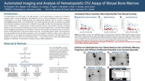 科学海报Automated Imaging and Analysis of Hematopoietic CFU Assays of Mouse Bone Marrow

 沪公网安备31010102008431号