Bone Marrow Niches and HSC Fates
A detailed reference on signaling pathways in the bone marrow and how these influence HSC fate decisions; created in partnership with Nature Reviews Immunology and Nature Reviews Molecular Cell Biology
Orelio C et al. (DEC 2008)
Blood 112 13 4895--904
Interleukin-1-mediated hematopoietic cell regulation in the aorta-gonad-mesonephros region of the mouse embryo.
Hematopoiesis during development is a dynamic process,with many factors involved in the emergence and regulation of hematopoietic stem cells (HSCs) and progenitor cells. Whereas previous studies have focused on developmental signaling and transcription factors in embryonic hematopoiesis,the role of well-known adult hematopoietic cytokines in the embryonic hematopoietic system has been largely unexplored. The cytokine interleukin-1 (IL-1),best known for its proinflammatory properties,has radioprotective effects on adult bone marrow HSCs,induces HSC mobilization,and increases HSC proliferation and/or differentiation. Here we examine IL-1 and its possible role in regulating hematopoiesis in the midgestation mouse embryo. We show that IL-1,IL-1 receptors (IL-1Rs),and signaling mediators are expressed in the aorta-gonad-mesonephros (AGM) region during the time when HSCs emerge in this site. IL-1 signaling is functional in the AGM,and the IL-1RI is expressed ventrally in the aortic subregion by some hematopoietic,endothelial,and mesenchymal cells. In vivo analyses of IL-1RI-deficient embryos show an increased myeloid differentiation,concomitant with a slight decrease in AGM HSC activity. Our results suggest that IL-1 is an important homeostatic regulator at the earliest time of HSC development,acting to limit the differentiation of some HSCs along the myeloid lineage.
View Publication
产品号#:
03434
03444
产品名:
MethoCult™ GF M3434
MethoCult™ GF M3434
Zhao H et al. (JAN 2009)
Blood 113 3 505--16
The c-myb proto-oncogene and microRNA-15a comprise an active autoregulatory feedback loop in human hematopoietic cells.
The c-myb proto-oncogene encodes an obligate hematopoietic cell transcription factor important for lineage commitment,proliferation,and differentiation. Given its critical functions,c-Myb regulatory factors are of great interest but remain incompletely defined. Herein we show that c-Myb expression is subject to posttranscriptional regulation by microRNA (miRNA)-15a. Using a luciferase reporter assay,we found that miR-15a directly binds the 3'-UTR of c-myb mRNA. By transfecting K562 myeloid leukemia cells with a miR-15a mimic,functionality of binding was shown. The mimic decreased c-Myb expression,and blocked the cells in the G(1) phase of cell cycle. Exogenous expression of c-myb mRNA lacking the 3'-UTR partially rescued the miR-15a induced cell-cycle block. Of interest,the miR-15a promoter contained several potential c-Myb protein binding sites. Occupancy of one canonical c-Myb binding site was demonstrated by chromatin immunoprecipitation analysis and shown to be required for miR-15a expression in K562 cells. Finally,in studies using normal human CD34(+) cells,we showed that c-Myb and miR-15a expression were inversely correlated in cells undergoing erythroid differentiation,and that overexpression of miR-15a blocked both erythroid and myeloid colony formation in vitro. In aggregate,these findings suggest the presence of a c-Myb-miR-15a autoregulatory feedback loop of potential importance in human hematopoiesis.
View Publication
产品号#:
09500
产品名:
BIT 9500血清替代物
Singh KP et al. (JAN 2009)
Carcinogenesis 30 1 11--9
Treatment of mice with the Ah receptor agonist and human carcinogen dioxin results in altered numbers and function of hematopoietic stem cells.
The aryl hydrocarbon receptor (AhR) mediates the carcinogenicity of a family of environmental contaminants,the most potent being 2,3,7,8-tetrachlorodibenzo-p-dioxin (TCDD). Increased incidence of lymphoma and leukemia in humans is associated with TCDD exposure. Although AhR activation by TCDD has profound effects on the immune system,precise cellular and molecular mechanisms have yet to be determined. These studies tested the hypothesis that alteration of marrow populations following treatment of mice with TCDD is due to an effect on hematopoietic stem cells (HSCs). Treatment with TCDD resulted in an increased number and proliferation of bone marrow (BM) populations enriched for HSCs. There was a time-dependent decrease in B-lineage cells with a concomitant increase in myeloid populations. The decrease in the B-cell lineage colony-forming unit-preB progenitors along with a transient increase in myeloid progenitors were consistent with a skewing of lineage development from lymphoid to myeloid populations. However,HSCs from TCDD-treated mice exhibited diminished capacity to reconstitute and home to marrow of irradiated recipients. AhR messenger RNA was expressed in progenitor subsets but is downregulated during HSC proliferation. This result was consistent with the lack of response following the exposure of 5-fluorouracil-treated mice to TCDD. The direct exposure of cultured BM cells to TCDD inhibited the growth of immature hematopoietic progenitor cells,but not more mature lineage-restricted progenitors. Overall,these data are consistent with the hypothesis that TCDD,through AhR activation,alters the ability of HSCs to respond appropriately to signals within the marrow microenvironment.
View Publication
产品号#:
03231
产品名:
MethoCult™ M3231
Matsumoto K et al. (JAN 2000)
Stem cells (Dayton,Ohio) 18 3 196--203
In vitro proliferation potential of AC133 positive cells in peripheral blood.
AC133 antigen is a novel marker for human hematopoietic stem/progenitor cells. In this study,we examined the expression and proliferation potential of AC133(+) cells obtained from steady-state peripheral blood (PB). The proportion of AC133(+) cells in the CD34(+) subpopulation of steady-state PB was significantly lower than that of cord blood (CB),although that of cytokine-mobilized PB was higher than that of CB. The proliferation potential of AC133(+)CD34(+) and AC133(-)CD34(+) cells was examined by colony-forming analysis and analysis of long-term culture-initiating cells (LTC-IC). Although the total number of colony-forming cells was essentially the same in the AC133(+)CD34(+) fraction as in the AC133(-)CD34(+) fraction,the proportion of LTC-IC was much higher in the AC133(+)CD34(+) fraction. Virtually no LTC-IC were detected in the AC133(-)CD34(+) fraction. In addition,the features of the colonies grown from these two fractions were quite different. Approximately 70% of the colonies derived from the AC133(+)CD34(+) fraction were granulocyte-macrophage colonies,whereas more than 90% of the colonies derived from the AC133(-)CD34(+) fraction were erythroid colonies. Furthermore,an ex vivo expansion study observed expansion of colony-forming cells only in the AC133(+)CD34(+) population,and not in the AC133(-)CD34(+) population. These findings suggest that to isolate primitive hematopoietic cells from steady-state PB,selection by AC133 expression is better than selection by CD34 expression.
View Publication
Haniffa M et al. (FEB 2009)
The Journal of experimental medicine 206 2 371--85
Differential rates of replacement of human dermal dendritic cells and macrophages during hematopoietic stem cell transplantation.
Animal models of hematopoietic stem cell transplantation have been used to analyze the turnover of bone marrow-derived cells and to demonstrate the critical role of recipient antigen-presenting cells (APC) in graft versus host disease (GVHD). In humans,the phenotype and lineage relationships of myeloid-derived tissue APC remain incompletely understood. It has also been proposed that the risk of acute GVHD,which extends over many months,is related to the protracted survival of certain recipient APC. Human dermis contains three principal subsets of CD45(+)HLA-DR(+) cells: CD1a(+)CD14(-) DC,CD1a(-)CD14(+) DC,and CD1a(-)CD14(+)FXIIIa(+) macrophages. In vitro,each subset has characteristic properties. After transplantation,both CD1a(+) and CD14(+) DC are rapidly depleted and replaced by donor cells,but recipient macrophages can be found in GVHD lesions and may persist for many months. Macrophages isolated from normal dermis secrete proinflammatory cytokines. Although they stimulate little proliferation of naive or memory CD4(+) T cells,macrophages induce cytokine expression in memory CD4(+) T cells and activation and proliferation of CD8(+) T cells. These observations suggest that dermal macrophages and DC are from distinct lineages and that persistent recipient macrophages,although unlikely to initiate alloreactivity,may contribute to GVHD by sustaining the responses of previously activated T cells.
View Publication
产品号#:
19155
19155RF
产品名:
Chan G et al. (APR 2009)
Blood 113 18 4414--24
Leukemogenic Ptpn11 causes fatal myeloproliferative disorder via cell-autonomous effects on multiple stages of hematopoiesis.
PTPN11,which encodes the tyrosine phosphatase SHP2,is mutated in approximately 35% of patients with juvenile myelomonocytic leukemia (JMML) and at a lower incidence in other neoplasms. To model JMML pathogenesis,we generated knockin mice that conditionally express the leukemia-associated mutant Ptpn11(D61Y). Expression of Ptpn11(D61Y) in all hematopoietic cells evokes a fatal myeloproliferative disorder (MPD),featuring leukocytosis,anemia,hepatosplenomegaly,and factor-independent colony formation by bone marrow (BM) and spleen cells. The Lin(-)Sca1(+)cKit(+) (LSK) compartment is expanded and right-shifted�
View Publication
产品号#:
03234
03334
03434
03444
产品名:
MethoCult™ M3234
MethoCult™ M3334
MethoCult™ GF M3434
MethoCult™ GF M3434
Pendino F et al. (APR 2009)
Blood 113 14 3172--81
Functional involvement of RINF, retinoid-inducible nuclear factor (CXXC5), in normal and tumoral human myelopoiesis.
Retinoids triggers differentiation of acute promyelocytic leukemia (APL) blasts by transcriptional regulation of myeloid regulatory genes. Using a microarray approach,we have identified a novel retinoid-responsive gene (CXXC5) encoding a nuclear factor,retinoid-inducible nuclear factor (RINF),that contains a CXXC-type zinc-finger motif. RINF expression correlates with retinoid-induced differentiation of leukemic cells and with cytokine-induced myelopoiesis of normal CD34(+) progenitors. Furthermore,short hairpin RNA (shRNA) interference suggests for this gene a regulatory function in both normal and tumoral myelopoiesis. Interestingly,RINF localizes to 5q31.3,a small region often deleted in myeloid leukemia (acute myeloid leukemia [AML]/myelodysplasia [MDS]) and suspected to harbor one or several tumor suppressor gene.
View Publication
产品号#:
70002
70002.1
70002.2
70002.3
70002.4
70002.5
产品名:
Povsic TJ et al. (OCT 2009)
Journal of thrombosis and thrombolysis 28 3 259--65
BACKGROUND: Interest in the biology of endogenous progenitor cells (EPCs) continues to grow as evidence of their role in vascular repair mounts. EPC enumeration requires specialized laboratory techniques and is performed immediately after sample acquisition,limiting the clinical contexts in which EPC enumeration can be performed and the ability to increase sample sizes through multi-center participation. METHODS: We compared the numbers of EPCs enumerated in samples processed immediately after acquisition (n = 36) with EPCs enumerated in specimens stored for 24 hours or after cryopreservation of mononuclear cells (MNC) using two EPC identification strategies: cell surface marker expression (CD133/CD34) and aldehyde dehydrogenase activity (ALDH(br) cells). RESULTS: EPCs assessed in fresh samples correlated with EPCs enumerated after whole blood storage (r = 0.699 for CD133(+)CD34(+) cells,r = 0.880 for ALDH(br) cells,P textless 0.005 and P textless 0.0001,respectively) or mononuclear cryopreservation (r = 0.590 for CD133(+)CD34(+) cells,r = 0.894 for ALDH(br) cells,P textless 0.0001 for each); however,correlation based on assessment of ALDH(br) cells was higher (P textless 0.0003 for comparison of correlation coefficients). Initial results from a multi-site clinical trial suggest that EPC enumeration after mononuclear cell cryopreservation is feasible. CONCLUSION: EPC analysis based on ALDH activity is reproducible,even after extended whole blood storage or MNC cryopreservation.
View Publication
产品号#:
01700
01705
01701
01702
产品名:
ALDEFLUOR™ 试剂盒
ALDEFLUOR™ DEAB试剂, 1.5 mM, 1 mL
ALDEFLUOR™检测缓冲液
Ali N et al. (APR 2009)
Blood 113 16 3690--5
Forward RNAi screens in primary human hematopoietic stem/progenitor cells.
The mechanisms regulating key fate decisions such as self-renewal and differentiation in hematopoietic stem and progenitor cells (HSPC) remain poorly understood. We report here a screening strategy developed to assess modulators of human hematopoiesis using a lentiviral short hairpin RNA (shRNA) library transduced into cord blood-derived stem/progenitor cells. To screen for modifiers of self-renewal/differentiation,we used the limited persistence of HSPCs under ex vivo culture conditions as a baseline for functional selection of shRNAs conferring enhanced maintenance or expansion of the stem/progenitor potential. This approach enables complex,pooled screens in large numbers of cells. Functional selection identified novel specific gene targets (exostoses 1) or shRNA constructs capable of altering human hematopoietic progenitor differentiation or stem cell expansion,respectively,thereby demonstrating the potential of this forward screening approach in primary human stem cell populations.
View Publication
产品号#:
09600
09650
产品名:
StemSpan™ SFEM
StemSpan™ SFEM
Orelio C et al. (APR 2009)
Haematologica 94 4 462--9
Interleukin-1 regulates hematopoietic progenitor and stem cells in the midgestation mouse fetal liver.
BACKGROUND: Hematopoietic progenitors are generated in the yolk sac and aorta-gonad-mesonephros region during early mouse development. At embryonic day 10.5 the first hematopoietic stem cells emerge in the aorta-gonad-mesonephros. Subsequently,hematopoietic stem cells and progenitors are found in the fetal liver. The fetal liver is a potent hematopoietic site,playing an important role in the expansion and differentiation of hematopoietic progenitors and hematopoietic stem cells. However,little is known concerning the regulation of fetal liver hematopoietic stem cells. In particular,the role of cytokines such as interleukin-1 in the regulation of hematopoietic stem cells in the embryo has been largely unexplored. Recently,we observed that the adult pro-inflammatory cytokine interleukin-1 is involved in regulating aorta-gonad-mesonephros hematopoietic progenitor and hematopoietic stem cell activity. Therefore,we set out to investigate whether interleukin-1 also plays a role in regulating fetal liver progenitor cells and hematopoietic stem cells. DESIGN AND METHODS: We examined the interleukin-1 ligand and receptor expression pattern in the fetal liver. The effects of interleukin-1 on hematopoietic progenitor cells and hematopoietic stem cells were studied by FACS and transplantation analyses of fetal liver explants,and in vivo effects on hematopoietic stem cell and progenitors were studied in Il1r1(-/-) embryos. RESULTS: We show that fetal liver hematopoietic progenitor cells express the IL-1RI and that interleukin-1 increases fetal liver hematopoiesis,progenitor cell activity and promotes hematopoietic cell survival. Moreover,we show that in Il1r1(-/-) embryos,hematopoietic stem cell activity is impaired and myeloid progenitor activity is increased. CONCLUSIONS: The IL-1 ligand and receptor are expressed in the midgestation liver and act in the physiological regulation of fetal liver hematopoietic progenitor cells and hematopoietic stem cells.
View Publication

 EasySep™小鼠TIL(CD45)正选试剂盒


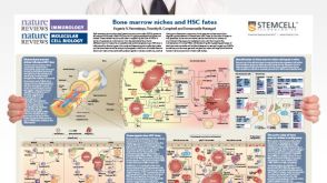 挂图Bone Marrow Niches and HSC Fates A detailed reference on signaling pathways in the bone marrow and how these influence HSC fate decisions; created in partnership with Nature Reviews Immunology and Nature Reviews Molecular Cell Biology
 技术窍门人造血干细胞和祖细胞表型的鉴定
 沪公网安备31010102008431号