Tauchmanovà et al. (MAY 2003)
Cancer 97 10 2453--61
Avascular necrosis in long-term survivors after allogeneic or autologous stem cell transplantation: a single center experience and a review.
BACKGROUND: The most debilitating skeletal complication of stem cell transplantation (SCT) is avascular necrosis (AVN). METHODS: Two hundred seven consecutive patients were evaluated prospectively for AVN. They survived disease free for more than 180 days after autologous or allogeneic SCT for hematologic malignancies. The diagnosis of AVN in suspicious cases was confirmed by magnetic resonance imaging. Possible correlations with treatments,bone mineral density (BMD),graft versus host disease (GVHD),and in vitro growth of fibroblast progenitors were investigated. Bone mineral density was evaluated by dual-energy X-ray absorptiometry in 100 transplanted patients,and the in vitro growth of fibroblast progenitors was monitored by a fibroblast colony-forming unit (CFU-F) assay in 30 patients after allogeneic SCT. RESULTS: Twelve patients developed AVN 3-114 months (median,26 months) following SCT: 10 (10%) after allogeneic SCT and 2 (1.9%) after autologous SCT (P = 0.04). Twenty-five joints were affected by AVN. All patients had femoral head involvement,which was managed with hip replacement in six of them. All but one patient who developed AVN after allogeneic SCT suffered from chronic GVHD (cGVHD). Avascular necrosis occurred 1-4 months after exacerbation or progression of cGVHD. Cumulative dose of steroids was similar in both SCT groups (including steroids given pretransplant for the basic disease),whereas treatment duration was significantly longer in the allogeneic SCT group. Avascular necrosis was related to the decreased number of bone marrow CFU-F colonies in vitro,but not to BMD values. CONCLUSIONS: Avascular necrosis is a skeletal complication that occurs more often after allogeneic than after autologous SCT. Occurrence of AVN symptoms after clinical follow-up of cGVHD suggests that cGVHD requiring long-term steroid therapy is one of the main risk factors for AVN. Avascular necrosis may be facilitated by a severe deficit in the repopulating capacity of bone marrow stromal stem cells after SCT.
View Publication
产品号#:
05401
05402
05411
产品名:
MesenCult™ MSC 基础培养基(人)
MesenCult™ MSC 刺激补充剂(人)
MesenCult™ 增殖试剂盒(人)
Rodrí et al. (MAY 2004)
Blood 103 9 3349--54
Interleukin-6 deficiency affects bone marrow stromal precursors, resulting in defective hematopoietic support.
Interleukin-6 (IL-6) is a critical factor in the regulation of stromal function and hematopoiesis. In vivo bromodeoxyuridine incorporation analysis indicates that the percentage of Lin(-)Sca-1(+) hematopoietic progenitors undergoing DNA synthesis is diminished in IL-6-deficient (IL-6(-/-)) bone marrow (BM) compared with wild-type BM. Reduced proliferation of IL-6(-/-) BM progenitors is also observed in IL-6(-/-) long-term BM cultures,which show defective hematopoietic support as measured by production of total cells,granulocyte macrophage-colony-forming units (CFU-GMs),and erythroid burst-forming units (BFU-Es). Seeding experiments of wild-type and IL-6(-/-) BM cells on irradiated wild-type or IL-6-deficient stroma indicate that the hematopoietic defect can be attributed to the stromal and not to the hematopoietic component. In IL-6(-/-) BM,stromal mesenchymal precursors,fibroblast CFUs (CFU-Fs),and stroma-initiating cells (SICs) are reduced to almost 50% of the wild-type BM value. Moreover,IL-6(-/-) stromata show increased CD34 and CD49e expression and reduced expression of the membrane antigens vascular cell adhesion molecule-1 (VCAM-1),Sca-1,CD49f,and Thy1. These data strongly suggest that IL-6 is an in vivo growth factor for mesenchymal precursors,which are in part implicated in the reduced longevity of the long-term repopulating stem cell compartment of IL-6(-/-) mice.
View Publication
产品号#:
03534
05501
05502
05350
28600
产品名:
MethoCult™ GF M3534
L-Calc™有限稀释软件
Bieback K et al. (JAN 2004)
Stem cells (Dayton,Ohio) 22 4 625--34
Critical parameters for the isolation of mesenchymal stem cells from umbilical cord blood.
Evidence has emerged that mesenchymal stem cells (MSCs) represent a promising population for supporting new clinical concepts in cellular therapy. However,attempts to isolate MSCs from umbilical cord blood (UCB) of full-term deliveries have previously either failed or been characterized by a low yield. We investigated whether cells with MSC characteristics and multi-lineage differentiation potential can be cultivated from UCB of healthy newborns and whether yields might be maximized by optimal culture conditions or by defining UCB quality criteria. Using optimized isolation and culture conditions,in up to 63% of 59 low-volume UCB units,cells showing a characteristic mesenchymal morphology and immune phenotype (MSC-like cells) were isolated. These were similar to control MSCs from adult bone marrow (BM). The frequency of MSC-like cells ranged from 0 to 2.3 clones per 1 x 10(8) mononuclear cells (MNCs). The cell clones proliferated extensively with at least 20 population doublings within eight passages. In addition,osteogenic and chondrogenic differentiation demonstrated a multi-lineage capacity comparable with BM MSCs. However,in contrast to MSCs,MSC-like cells showed a reduced sensitivity to undergo adipogenic differentiation. Crucial points to isolate MSC-like cells from UCB were a time from collection to isolation of less than 15 hours,a net volume of more than 33 ml,and an MNC count of more than 1 x 10(8) MNCs. Because MSC-like cells can be isolated at high efficacy from full-term UCB donations,we regard UCB as an additional stem cell source for experimental and potentially clinical purposes.
View Publication
产品号#:
05401
05402
05411
产品名:
MesenCult™ MSC 基础培养基(人)
MesenCult™ MSC 刺激补充剂(人)
MesenCult™ 增殖试剂盒(人)
Lee J-H et al. (JUL 2005)
Experimental cell research 307 1 174--82
Contribution of human bone marrow stem cells to individual skeletal myotubes followed by myogenic gene activation.
Much attention is focused on characterizing the contribution of bone marrow (BM)-derived cells to regenerating skeletal muscle,fuelled by hopes for stem cell-mediated therapy of muscle degenerative diseases. Though physical integration of BM stem cells has been well documented,little evidence of functional commitment to myotube phenotype has been reported. This is due to the innate difficulty in distinguishing gene products derived from donor versus host nuclei. Here,we demonstrate that BM-derived stem cells contribute via gene expression following incorporation to skeletal myotubes. By co-culturing human BM-derived mesenchymal stem cells (MSC) with mouse skeletal myoblasts,physical incorporation was observed by genetic lineage tracing and species-specific immunofluorescence. We used a human-specific antibody against the intermediate filament protein nestin,a marker of regenerating skeletal muscle,to identify functional contribution of MSC to myotube formation. Although nestin expression was never detected in MSC,human-specific expression was detected in myotubes that also contained MSC-derived nuclei. This induction of gene expression following myotube integration suggests that bone marrow-derived stem cells can reprogram and functionally contribute to the muscle cell phenotype. We propose that this model of myogenic commitment may provide the means to further characterize functional reprogramming of MSC to skeletal muscle.
View Publication
产品号#:
05401
15128
15168
产品名:
MesenCult™ MSC 基础培养基(人)
RosetteSep™人间充质干细胞富集抗体混合物
RosetteSep™人间充质干细胞富集抗体混合物
Zhang H et al. (NOV 2005)
American journal of physiology. Heart and circulatory physiology 289 5 H2089--96
Increasing donor age adversely impacts beneficial effects of bone marrow but not smooth muscle myocardial cell therapy.
We evaluated the impact of donor age on the efficacy of myocardial cellular therapy for ischemic cardiomyopathy. Characteristics of smooth muscle cells (SMC),bone marrow stromal cells (MSCs),and skeletal muscle cells (SKMCs) from young,adult,and old rats were compared in vitro. Three weeks after coronary ligation,3.5 million SMCs (n = 11) or MSCs (n = 9) from old syngenic rats or culture medium (n = 6) were injected into the ischemic region. Five weeks after implantation,cardiac function was assessed by echocardiography and the Langendorff apparatus. In the in vitro study,the numbers and proliferation of MSCs from fresh bone marrow and SKMCs from fresh tissue but not SMCs were markedly diminished in old animals (P textless 0.05 both groups). SKMCs from old animals did not reach confluence. After treatment with 5-azacytidine (azacitidine),the myogenic potential of old MSCs was decreased compared with young MSCs. In the in vivo study,both SMC and MSC transplantation induced significant angiogenesis compared with media injections (P textless 0.05 both groups). Transplantation of SMCs but not MSCs prevented scar thinning (P = 0.03) and improved ejection fraction and fractional shortening (P textless 0.05). Load-independent indices of cardiac function in a Langendorff preparation confirmed improved function in the aged SMC group (P = 0.01) but not in the MSC group compared with the control group. In conclusion,donor age adversely impacts the efficacy of cellular therapy for myocardial regeneration and is cell-type dependent. SMCs from old donors retain their ability to improve cardiac function after implantation into ischemic myocardium.
View Publication
产品号#:
05501
05502
产品名:
Muguruma Y et al. (MAR 2006)
Blood 107 5 1878--87
Reconstitution of the functional human hematopoietic microenvironment derived from human mesenchymal stem cells in the murine bone marrow compartment.
Hematopoiesis is maintained by specific interactions between both hematopoietic and nonhematopoietic cells. Whereas hematopoietic stem cells (HSCs) have been extensively studied both in vitro and in vivo,little is known about the in vivo characteristics of stem cells of the nonhematopoietic component,known as mesenchymal stem cells (MSCs). Here we have visualized and characterized human MSCs in vivo following intramedullary transplantation of enhanced green fluorescent protein-marked human MSCs (eGFP-MSCs) into the bone marrow (BM) of nonobese diabetic/severe combined immunodeficiency (NOD/SCID) mice. Between 4 to 10 weeks after transplantation,eGFP-MSCs that engrafted in murine BM integrated into the hematopoietic microenvironment (HME) of the host mouse. They differentiated into pericytes,myofibroblasts,BM stromal cells,osteocytes in bone,bone-lining osteoblasts,and endothelial cells,which constituted the functional components of the BM HME. The presence of human MSCs in murine BM resulted in an increase in functionally and phenotypically primitive human hematopoietic cells. Human MSC-derived cells that reconstituted the HME appeared to contribute to the maintenance of human hematopoiesis by actively interacting with primitive human hematopoietic cells.
View Publication
产品号#:
04034
04044
产品名:
MethoCult™ H4034 Optimum
MethoCult™ H4034 Optimum
Maes C et al. (MAY 2006)
The Journal of clinical investigation 116 5 1230--42
Placental growth factor mediates mesenchymal cell development, cartilage turnover, and bone remodeling during fracture repair.
Current therapies for delayed- or nonunion bone fractures are still largely ineffective. Previous studies indicated that the VEGF homolog placental growth factor (PlGF) has a more significant role in disease than in health. Therefore we investigated the role of PlGF in a model of semi-stabilized bone fracture healing. Fracture repair in mice lacking PlGF was impaired and characterized by a massive accumulation of cartilage in the callus,reminiscent of delayed- or nonunion fractures. PlGF was required for the early recruitment of inflammatory cells and the vascularization of the fracture wound. Interestingly,however,PlGF also played a role in the subsequent stages of the repair process. Indeed in vivo and in vitro findings indicated that PlGF induced the proliferation and osteogenic differentiation of mesenchymal progenitors and stimulated cartilage turnover by particular MMPs. Later in the process,PlGF was required for the remodeling of the newly formed bone by stimulating osteoclast differentiation. As PlGF expression was increased throughout the process of bone repair and all the important cell types involved expressed its receptor VEGFR-1,the present data suggest that PlGF is required for mediating and coordinating the key aspects of fracture repair. Therefore PlGF may potentially offer therapeutic advantages for fracture repair.
View Publication
产品号#:
03534
03334
03434
03444
18753
18753RF
产品名:
MethoCult™ GF M3534
MethoCult™ M3334
MethoCult™ GF M3434
MethoCult™ GF M3434
Lapter S et al. (MAR 2007)
Stem cells (Dayton,Ohio) 25 3 761--70
Structure and implied functions of truncated B-cell receptor mRNAs in early embryo and adult mesenchymal stem cells: Cdelta replaces Cmu in mu heavy chain-deficient mice.
Stem cells exhibit a promiscuous gene expression pattern. We show herein that the early embryo and adult MSCs express B-cell receptor component mRNAs. To examine possible bearings of these genes on the expressing cells,we studied immunoglobulin mu chain-deficient mice. Pregnant mu chain-deficient females were found to produce a higher percentage of defective morulae compared with control females. Structure analysis indicated that the mu mRNA species found in embryos and in mesenchyme consist of the constant region of the mu heavy chain that encodes a recombinant 50-kDa protein. In situ hybridization localized the constant mu gene expression to loose mesenchymal tissues within the day-12.5 embryo proper and the yolk sac. In early embryo and in adult mesenchyme from mu-deficient mice,delta replaced mu chain,implying a possible requirement of these alternative molecules for embryo development and mesenchymal functions. Indeed,overexpression of the mesenchymal-truncated mu heavy chain in 293T cells resulted in specific subcellular localization and in G(1) growth arrest. The lack of such occurrence following overexpression of a complete,rearranged form of mu chain suggests that the mesenchymal version of this mRNA may possess unique functions.
View Publication
产品号#:
05501
05502
产品名:
Seeger FH et al. (MAR 2007)
European heart journal 28 6 766--72
Cell isolation procedures matter: a comparison of different isolation protocols of bone marrow mononuclear cells used for cell therapy in patients with acute myocardial infarction.
AIM: The recently published REPAIR-AMI and ASTAMI trial showed differences in contractile recovery of left ventricular function after infusion of bone marrow-derived cells in acute myocardial infarction. Since the trials used different protocols for cell isolation and storage (REPAIR-AMI: Ficoll,storage in X-vivo 10 medium plus serum; ASTAMI: Lymphoprep,storage in NaCl plus plasma),we compared the functional activity of BMC isolated by the two different protocols. METHODS AND RESULTS: The recovery of total cell number,colony-forming units (CFU),and the number of mesenchymal stem cells were significantly reduced to 77 +/- 4%,83 +/- 16%,and 65 +/- 15%,respectively,when using the ASTAMI protocol compared with the REPAIR protocol. The capacity of the isolated BMC to migrate in response to stromal cell-derived factor 1 (SDF-1) was profoundly reduced when using the ASTAMI cell isolation procedure (42 +/- 8% and 78 +/- 3% reduction in healthy and CAD-patient cells,respectively). Finally,infusion of BMC into a hindlimb ischaemia model demonstrated a significantly blunted blood-flow-recovery by BMC isolated with the ASTAMI protocol (54 +/- 6% of the effect obtained by REPAIR cells). Comparison of the individual steps identified the use of NaCl and plasma for cell storage as major factors for functional impairment of the BMC. CONCLUSION: Cell isolation protocols have a major impact on the functional activity of bone marrow-derived progenitor cells. The assessment of cell number and viability may not entirely reflect the functional capacity of cells in vivo. Additional functional testing appears to be mandatory to assure proper cell function before embarking on clinical cell therapy trials.
View Publication

 EasySep™小鼠TIL(CD45)正选试剂盒


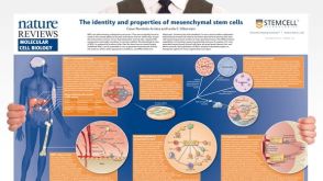 挂图The Identity and Properties of Mesenchymal Stem Cells Overview of MSC expansion, differentiation, immunoregulatory properties and therapeutic potential
 沪公网安备31010102008431号