LFA-1 activity state on dendritic cells regulates contact duration with T cells and promotes T-cell priming.
A key event in the successful induction of adaptive immune responses is the antigen-specific activation of T cells by dendritic cells (DCs). Although LFA-1 (lymphocyte function-associated antigen 1) on T cells is considered to be important for antigen-specific T-cell activation,the role for LFA-1 on DCs remains elusive. Using 2 different approaches to activate LFA-1 on DCs,either by deletion of the αL-integrin cytoplasmic GFFKR sequence or by silencing cytohesin-1-interacting protein,we now provide evidence that DCs are able to make use of active LFA-1 and can thereby control the contact duration with naive T cells. Enhanced duration of DC/T-cell interaction correlates inversely with antigen-specific T-cell proliferation,generation of T-helper 1 cells,and immune responses leading to delayed-type hypersensitivity. We could revert normal interaction time and T-cell proliferation to wild-type levels by inhibition of active LFA-1 on DCs. Our data further suggest that cytohesin-1-interacting protein might be responsible for controlling LFA-1 deactivation on mature DCs. In summary,our findings indicate that LFA-1 on DCs needs to be in an inactive state to ensure optimal T-cell activation and suggest that regulation of LFA-1 activity allows DCs to actively control antigen-driven T-cell proliferation and effective immune responses.
View Publication
产品号#:
21000
20119
20155
19752
19752RF
19753
19753RF
产品名:
RoboSep™- S
RoboSep™ 吸头组件抛光剂
RoboSep™分选管套装(9个塑料管)
Takeuchi H et al. (NOV 2010)
Journal of immunology (Baltimore,Md. : 1950) 185 9 5289--99
Efficient induction of CCR9 on T cells requires coactivation of retinoic acid receptors and retinoid X receptors (RXRs): exaggerated T Cell homing to the intestine by RXR activation with organotins.
The active vitamin A metabolite retinoic acid (RA) imprints gut-homing specificity on lymphocytes upon activation by inducing the expression of α4β7 integrin and CCR9. RA receptor (RAR) activation is essential for their expression,whereas retinoid X receptor (RXR) activation is not essential for α4β7 expression. However,it remains unclear whether RXR activation affects the RA-dependent CCR9 expression on T cells and their gut homing. The major physiological RA,all-trans-RA,binds to RAR but not to RXR at physiological concentrations. Cell-surface CCR9 expression was often induced on a limited population of murine naive CD4(+) T cells by all-trans-RA or the RAR agonist Am80 alone upon CD3/CD28-mediated activation in vitro,but it was markedly enhanced by adding the RXR agonist PA024 or the RXR-binding environmental chemicals tributyltin and triphenyltin. Accordingly,CD4(+) T cells treated with the combination of all-trans-RA and tributyltin migrated into the small intestine upon adoptive transfer much more efficiently than did those treated with all-trans-RA alone. Furthermore,naive TCR transgenic CD4(+) T cells transferred into wild-type recipients migrated into the small intestinal lamina propria following i.p. injection of Ag,and the migration was enhanced by i.p. injection of PA024. We also show that PA024 markedly enhanced the all-trans-RA-induced CCR9 expression on naturally occurring naive-like regulatory T cells upon activation,resulting in the expression of high levels of α4β7,CCR9,and Foxp3. These results suggest that RXR activation enhances the RAR-dependent expression of CCR9 on T cells and their homing capacity to the small intestine.
View Publication
产品号#:
19752
19752RF
产品名:
Katzman SD et al. (OCT 2010)
Proceedings of the National Academy of Sciences of the United States of America 107 42 18085--90
Duration of antigen receptor signaling determines T-cell tolerance or activation.
The early events that determine the decision between lymphocyte tolerance and activation are not well-understood. Using a model of systemic self-antigen recognition by CD4(+) T cells,we show,using single-cell biochemical analyses,that tolerance is characterized by transient signaling events downstream of T-cell receptor engagement in the mammalian target of rapamycin (mTOR) and NF-κB pathways. Parallel studies done by live cell imaging show that the key difference between tolerance and activation is the duration of the T cell-antigen presenting cell (APC) interaction,as revealed by stable T-cell immobilization on antigen encounter. Brief T cell-APC interactions result in tolerance,and prolonged interactions are associated with activation and the development of effector cells. These studies show that the duration of T cell-APC interactions and magnitude of associated TCR-mediated signaling are key determinants of lymphocyte tolerance vs. activation.
View Publication
CD80 and PD-L2 define functionally distinct memory B cell subsets that are independent of antibody isotype
Memory B cells (MBCs) are long-lived sources of rapid,isotype-switched secondary antibody-forming cell (AFC) responses. Whether MBCs homogeneously retain the ability to self-renew and terminally differentiate or if these functions are compartmentalized into MBC subsets has remained unclear. It has been suggested that antibody isotype controls MBC differentiation upon restimulation. Here we demonstrate that subcategorizing MBCs on the basis of their expression of CD80 and PD-L2,independently of isotype,identified MBC subsets with distinct functions upon rechallenge. CD80(+)PD-L2(+) MBCs differentiated rapidly into AFCs but did not generate germinal centers (GCs); conversely,CD80(-)PD-L2(-) MBCs generated few early AFCs but robustly seeded GCs. The gene-expression patterns of the subsets supported both the identity and function of these distinct MBC types. Hence,the differentiation and regeneration of MBCs are compartmentalized.
View Publication
产品号#:
19752
19752RF
19754
19754RF
产品名:
Chevalier MF et al. ( 2015)
The Journal of Infectious Diseases 211 5 769--779
Phenotype Alterations in Regulatory T-Cell Subsets in Primary HIV Infection and Identification of Tr1-like Cells as the Main Interleukin 10-Producing CD4+ T Cells
BACKGROUND: Conventional regulatory T cells (Tregs) can suppress human immunodeficiency virus type 1 (HIV-1)-specific immune responses but cannot control immune activation in primary HIV infection. Here,we characterized Treg subsets,using recently defined phenotypic delineation,and analyzed the relative contribution of cell subsets to the production of immunosuppressive cytokines in primary HIV infection. METHODS: In a longitudinal prospective study,ex vivo phenotyping of fresh peripheral blood mononuclear cells from patients with primary HIV infection was performed at baseline and month 6 of follow-up to characterize Treg subsets,immune activation,and cytokine production in isolated CD4(+) T cells. RESULTS: The frequency of CD4(+)CD25(+)CD127(low) Tregs and the distribution between the naive,memory,and activated/memory Treg subsets was similar in patients and healthy donors. However,Tregs from patients with primary HIV infection showed peculiar phenotypic profiles,such as elevated FoxP3,ICOS,and CTLA-4 expression,with CTLA-4 expression strikingly increased in all Treg subsets both at baseline and month 6 of follow-up. The great majority of interleukin 10 (IL-10)-producing CD4(+) T cells were FoxP3(neg) (ie,Tr1-like cells). In contrast to conventional Tregs,Tr1-like cells were inversely correlated with immune activation and not associated with lower effector T-cell responses. CONCLUSION: FoxP3(neg) Tr1-like cells-major contributors to IL-10 production-may have a beneficial role by controlling immune activation in early HIV infection.
View Publication
产品号#:
18062
18062RF
15022
15062
18251
18251RF
21000
20119
20155
15021
15061
产品名:
RosetteSep™人CD4+ T细胞富集抗体混合物
RosetteSep™人CD4+ T细胞富集抗体混合物
RoboSep™- S
RoboSep™ 吸头组件抛光剂
RoboSep™分选管套装(9个塑料管)
RosetteSep™人T细胞富集抗体混合物
RosetteSep™人T细胞富集抗体混合物
Nova-Lamperti E et al. (JAN 2016)
Scientific Reports 6 20044
IL-10-produced by human transitional B-cells down-regulates CD86 expression on B-cells leading to inhibition of CD4+T-cell responses.
A novel subset of human regulatory B-cells has recently been described. They arise from within the transitional B-cell subpopulation and are characterised by the production of IL-10. They appear to be of significant importance in regulating T-cell immunity in vivo. Despite this important function,the molecular mechanisms by which they control T-cell activation are incompletely defined. Here we show that transitional B-cells produced more IL-10 and expressed higher levels of IL-10 receptor after CD40 engagement compared to other B-cell subsets. Furthermore,under this stimulatory condition,CD86 expressed by transitional B-cells was down regulated and T-cell proliferation was reduced. We provide evidence to demonstrate that the down-regulation of CD86 expression by transitional B-cells was due to the autocrine effect of IL-10,which in turn leads to decreased T-cell proliferation and TNF-α production. This analysis was further extended to peripheral B-cells in kidney transplant recipients. We observed that B-cells from patients tolerant to the graft maintained higher IL-10 production after CD40 ligation,which correlates with lower CD86 expression compared to patients with chronic rejection. Hence,the results obtained in this study shed light on a new alternative mechanism by which transitional B-cells inhibit T-cell proliferation and cytokine production.
View Publication
产品号#:
15022
15062
15024
15064
产品名:
RosetteSep™人CD4+ T细胞富集抗体混合物
RosetteSep™人CD4+ T细胞富集抗体混合物
RosetteSep™人B细胞富集抗体混合物
RosetteSep™人B细胞富集抗体混合物
Thompson EA et al. (APR 2016)
Journal of Immunology 196 7 3054--63
Shortened Intervals during Heterologous Boosting Preserve Memory CD8 T Cell Function but Compromise Longevity.
Developing vaccine strategies to generate high numbers of Ag-specific CD8 T cells may be necessary for protection against recalcitrant pathogens. Heterologous prime-boost-boost immunization has been shown to result in large quantities of functional memory CD8 T cells with protective capacities and long-term stability. Completing the serial immunization steps for heterologous prime-boost-boost can be lengthy,leaving the host vulnerable for an extensive period of time during the vaccination process. We show in this study that shortening the intervals between boosting events to 2 wk results in high numbers of functional and protective Ag-specific CD8 T cells. This protection is comparable to that achieved with long-term boosting intervals. Short-boosted Ag-specific CD8 T cells display a canonical memory T cell signature associated with long-lived memory and have identical proliferative potential to long-boosted T cells Both populations robustly respond to antigenic re-exposure. Despite this,short-boosted Ag-specific CD8 T cells continue to contract gradually over time,which correlates to metabolic differences between short- and long-boosted CD8 T cells at early memory time points. Our studies indicate that shortening the interval between boosts can yield abundant,functional Ag-specific CD8 T cells that are poised for immediate protection; however,this is at the expense of forming stable long-term memory.
View Publication
Griffin DO et al. (JAN 2011)
The Journal of experimental medicine 208 1 67--80
Human B1 cells in umbilical cord and adult peripheral blood express the novel phenotype CD20+ CD27+ CD43+ CD70-.
B1 cells differ in many ways from conventional B cells,most prominently in the production of natural immunoglobulin,which is vitally important for protection against pathogens. B1 cells have also been implicated in the pathogenesis of autoimmune dyscrasias and malignant diseases. It has been impossible to accurately study B1 cells during health and illness because the nature of human B1 cells has not been successfully defined. This has produced controversy regarding the existence of human B1 cells. Here,we determined the phenotype of human B1 cells by testing sort-purified B cell fractions for three fundamental B1 cell functions based on mouse studies: spontaneous IgM secretion,efficient T cell stimulation,and tonic intracellular signaling. We found that a small population of CD20(+)CD27(+)CD43(+) cells present in both umbilical cord and adult peripheral blood fulfilled these criteria and expressed a skewed B cell receptor repertoire. These B cells express little or no surface CD69 and CD70,both of which are markedly up-regulated after activation of CD20(+)CD27(-)CD43(-) (naive) and CD20(+)CD27(+)CD43(-) (memory) B cells. This work identifies human B1 cells as CD20(+)CD27(+)CD43(+)CD70(-). We determined that the proportion of B1 cells declines with age,which may contribute to disease susceptibility. Identification of human B1 cells provides a foundation for future studies on the nature and role of these cells in human disease.
View Publication


 EasySep™小鼠TIL(CD45)正选试剂盒




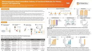 科学海报Mechanoporation-Based Intracellular Delivery of Functional Molecules for Primary Immune Cell Engineering

 沪公网安备31010102008431号