Ohoka Y et al. (JAN 2011)
Journal of immunology (Baltimore,Md. : 1950) 186 2 733--44
Retinoic acid-induced CCR9 expression requires transient TCR stimulation and cooperativity between NFATc2 and the retinoic acid receptor/retinoid X receptor complex.
Retinoic acid (RA) imprints gut-homing specificity on T cells upon activation by inducing the expression of chemokine receptor CCR9 and integrin α4β7. CCR9 expression seemed to be more highly dependent on RA than was the α4β7 expression,but its molecular mechanism remained unclear. In this article,we show that NFAT isoforms NFATc1 and NFATc2 directly interact with RA receptor (RAR) and retinoid X receptor (RXR) but play differential roles in RA-induced CCR9 expression on murine naive CD4(+) T cells. TCR stimulation for 6-24 h was required for the acquisition of responsiveness to RA and induced activation of NFATc1 and NFATc2. However,RA failed to induce CCR9 expression as long as TCR stimulation continued. After terminating TCR stimulation or adding cyclosporin A to the culture,Ccr9 gene transcription was induced,accompanied by inactivation of NFATc1 and sustained activation of NFATc2. Reporter and DNA-affinity precipitation assays demonstrated that the binding of NFATc2 to two NFAT-binding sites and that of the RAR/RXR complex to an RA response element half-site in the 5'-flanking region of the mouse Ccr9 gene were critical for RA-induced promoter activity. NFATc2 directly bound to RARα and RXRα,and it enhanced the binding of RARα to the RA response element half-site. NFATc1 also bound to the NFAT-binding sites and directly to RARα and RXRα,but it inhibited the NFATc2-dependent promoter activity. These results suggest that the cooperativity between NFATc2 and the RAR/RXR complex is essential for CCR9 expression on T cells and that NFATc1 interferes with the action of NFATc2.
View Publication
产品号#:
19752
19752RF
产品名:
Katzman SD et al. (OCT 2010)
Proceedings of the National Academy of Sciences of the United States of America 107 42 18085--90
Duration of antigen receptor signaling determines T-cell tolerance or activation.
The early events that determine the decision between lymphocyte tolerance and activation are not well-understood. Using a model of systemic self-antigen recognition by CD4(+) T cells,we show,using single-cell biochemical analyses,that tolerance is characterized by transient signaling events downstream of T-cell receptor engagement in the mammalian target of rapamycin (mTOR) and NF-κB pathways. Parallel studies done by live cell imaging show that the key difference between tolerance and activation is the duration of the T cell-antigen presenting cell (APC) interaction,as revealed by stable T-cell immobilization on antigen encounter. Brief T cell-APC interactions result in tolerance,and prolonged interactions are associated with activation and the development of effector cells. These studies show that the duration of T cell-APC interactions and magnitude of associated TCR-mediated signaling are key determinants of lymphocyte tolerance vs. activation.
View Publication
产品号#:
19752
19752RF
产品名:
Kieback E et al. (MAY 2016)
Immunity 44 5 1114--26
Thymus-Derived Regulatory T Cells Are Positively Selected on Natural Self-Antigen through Cognate Interactions of High Functional Avidity.
Regulatory T (Treg) cells expressing Foxp3 transcripton factor are essential for immune homeostasis. They arise in the thymus as a separate lineage from conventional CD4(+)Foxp3(-) T (Tconv) cells. Here,we show that the thymic development of Treg cells depends on the expression of their endogenous cognate self-antigen. The formation of these cells was impaired in mice lacking this self-antigen,while Tconv cell development was not negatively affected. Thymus-derived Treg cells were selected by self-antigens in a specific manner,while autoreactive Tconv cells were produced through degenerate recognition of distinct antigens. These distinct modes of development were associated with the expression of T cell receptor of higher functional avidity for self-antigen by Treg cells than Tconv cells,a difference subsequently essential for the control of autoimmunity. Our study documents how self-antigens define the repertoire of thymus-derived Treg cells to subsequently endow this cell type with the capacity to undermine autoimmune attack.
View Publication
产品号#:
18782
18782RF
19852
19852RF
产品名:
EasySep™小鼠CD25调节性T细胞正选试剂盒
RoboSep™ 小鼠CD25调节性T细胞正选试剂盒
EasySep™小鼠CD4+ T细胞分选试剂盒
RoboSep™ 小鼠CD4+ T细胞分选试剂盒
Albert BJ et al. (AUG 2017)
Scientific reports 7 1 7456
Combinations of isoform-targeted histone deacetylase inhibitors and bryostatin analogues display remarkable potency to activate latent HIV without global T-cell activation.
Current antiretroviral therapy (ART) for HIV/AIDS slows disease progression by reducing viral loads and increasing CD4 counts. Yet ART is not curative due to the persistence of CD4+ T-cell proviral reservoirs that chronically resupply active virus. Elimination of these reservoirs through the administration of synergistic combinations of latency reversing agents (LRAs),such as histone deacetylase (HDAC) inhibitors and protein kinase C (PKC) modulators,provides a promising strategy to reduce if not eradicate the viral reservoir. Here,we demonstrate that largazole and its analogues are isoform-targeted histone deacetylase inhibitors and potent LRAs. Significantly,these isoform-targeted HDAC inhibitors synergize with PKC modulators,namely bryostatin-1 analogues (bryologs). Implementation of this unprecedented LRA combination induces HIV-1 reactivation to unparalleled levels and avoids global T-cell activation within resting CD4+ T-cells.
View Publication
产品号#:
19052
19052RF
17861
产品名:
EasySep™人CD4+ T细胞富集试剂盒
RoboSep™ 人CD4+ T细胞富集试剂盒含滤芯吸头
EasySep™人Pan-CD25正选和去除试剂盒
Pekalski ML et al. (AUG 2017)
JCI insight 2 16
Neonatal and adult recent thymic emigrants produce IL-8 and express complement receptors CR1 and CR2.
The maintenance of peripheral naive T lymphocytes in humans is dependent on their homeostatic division,not continuing emigration from the thymus,which undergoes involution with age. However,postthymic maintenance of naive T cells is still poorly understood. Previously we reported that recent thymic emigrants (RTEs) are contained in CD31+CD25- naive T cells as defined by their levels of signal joint T cell receptor rearrangement excision circles (sjTRECs). Here,by differential gene expression analysis followed by protein expression and functional studies,we define that the naive T cells having divided the least since thymic emigration express complement receptors (CR1 and CR2) known to bind complement C3b- and C3d-decorated microbial products and,following activation,produce IL-8 (CXCL8),a major chemoattractant for neutrophils in bacterial defense. We also observed an IL-8-producing memory T cell subpopulation coexpressing CR1 and CR2 and with a gene expression signature resembling that of RTEs. The functions of CR1 and CR2 on T cells remain to be determined,but we note that CR2 is the receptor for Epstein-Barr virus,which is a cause of T cell lymphomas and a candidate environmental factor in autoimmune disease.
View Publication
Abadier M et al. (DEC 2017)
Cell reports 21 13 3885--3899
Effector and Regulatory T Cells Roll at High Shear Stress by Inducible Tether and Sling Formation.
The adaptive immune response involves T cell differentiation and migration to sites of inflammation. T cell trafficking is initiated by rolling on inflamed endothelium. Tethers and slings,discovered in neutrophils,facilitate cell rolling at high shear stress. Here,we demonstrate that the ability to form tethers and slings during rolling is highly inducible in T helper 1 (Th1),Th17,and regulatory T (Treg) cells but less in Th2 cells. In vivo,endogenous Treg cells rolled stably in cremaster venules at physiological shear stress. Quantitative dynamic footprinting nanoscopy of Th1,Th17,and Treg cells uncovered the formation of multiple tethers per cell. Human Th1 cells also showed tethers and slings. RNA sequencing (RNA-seq) revealed the induction of cell migration and cytoskeletal genes in sling-forming cells. We conclude that differentiated CD4 T cells stabilize rolling by inducible tether and sling formation. These phenotypic changes approximate the adhesion phenotype of neutrophils and support CD4 T cell access to sites of inflammation.
View Publication
产品号#:
19762
19762RF
产品名:
EasySep™小鼠中性粒细胞富集试剂盒
RoboSep™ 小鼠中性粒细胞富集试剂盒含滤芯吸头
Balkow S et al. (SEP 2010)
Blood 116 11 1885--94
LFA-1 activity state on dendritic cells regulates contact duration with T cells and promotes T-cell priming.
A key event in the successful induction of adaptive immune responses is the antigen-specific activation of T cells by dendritic cells (DCs). Although LFA-1 (lymphocyte function-associated antigen 1) on T cells is considered to be important for antigen-specific T-cell activation,the role for LFA-1 on DCs remains elusive. Using 2 different approaches to activate LFA-1 on DCs,either by deletion of the αL-integrin cytoplasmic GFFKR sequence or by silencing cytohesin-1-interacting protein,we now provide evidence that DCs are able to make use of active LFA-1 and can thereby control the contact duration with naive T cells. Enhanced duration of DC/T-cell interaction correlates inversely with antigen-specific T-cell proliferation,generation of T-helper 1 cells,and immune responses leading to delayed-type hypersensitivity. We could revert normal interaction time and T-cell proliferation to wild-type levels by inhibition of active LFA-1 on DCs. Our data further suggest that cytohesin-1-interacting protein might be responsible for controlling LFA-1 deactivation on mature DCs. In summary,our findings indicate that LFA-1 on DCs needs to be in an inactive state to ensure optimal T-cell activation and suggest that regulation of LFA-1 activity allows DCs to actively control antigen-driven T-cell proliferation and effective immune responses.
View Publication

 EasySep™小鼠TIL(CD45)正选试剂盒


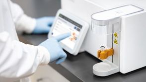 实验方案Optimizing Delivery Efficiency with Fluorescent Dextran Using the CellPore™ Transfection System
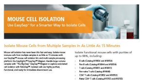
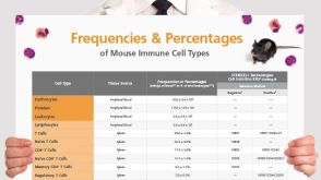 挂图Frequencies and Percentages of Mouse Immune Cell Types List of the frequencies of over 25 immune cell types in C57BL/6 mice


 沪公网安备31010102008431号