Carmona-Mora P et al. (OCT 2015)
Human Genetics 134 10 1099--1115
The nuclear localization pattern and interaction partners of GTF2IRD1 demonstrate a role in chromatin regulation
GTF2IRD1 is one of the three members of the GTF2I gene family,clustered on chromosome 7 within a 1.8 Mb region that is prone to duplications and deletions in humans. Hemizygous deletions cause Williams-Beuren syndrome (WBS) and duplications cause WBS duplication syndrome. These copy number variations disturb a variety of developmental systems and neurological functions. Human mapping data and analyses of knockout mice show that GTF2IRD1 and GTF2I underpin the craniofacial abnormalities,mental retardation,visuospatial deficits and hypersociability of WBS. However,the cellular role of the GTF2IRD1 protein is poorly understood due to its very low abundance and a paucity of reagents. Here,for the first time,we show that endogenous GTF2IRD1 has a punctate pattern in the nuclei of cultured human cell lines and neurons. To probe the functional relationships of GTF2IRD1 in an unbiased manner,yeast two-hybrid libraries were screened,isolating 38 novel interaction partners,which were validated in mammalian cell lines. These relationships illustrate GTF2IRD1 function,as the isolated partners are mostly involved in chromatin modification and transcriptional regulation,whilst others indicate an unexpected role in connection with the primary cilium. Mapping of the sites of protein interaction also indicates key features regarding the evolution of the GTF2IRD1 protein. These data provide a visual and molecular basis for GTF2IRD1 nuclear function that will lead to an understanding of its role in brain,behaviour and human disease.
View Publication
产品号#:
05850
05857
05870
05875
85850
85857
85870
85875
产品名:
mTeSR™1
mTeSR™1
Duan S et al. (DEC 2015)
Nature communications 6 10068
PTEN deficiency reprogrammes human neural stem cells towards a glioblastoma stem cell-like phenotype.
PTEN is a tumour suppressor frequently mutated in many types of cancers. Here we show that targeted disruption of PTEN leads to neoplastic transformation of human neural stem cells (NSCs),but not mesenchymal stem cells. PTEN-deficient NSCs display neoplasm-associated metabolic and gene expression profiles and generate intracranial tumours in immunodeficient mice. PTEN is localized to the nucleus in NSCs,binds to the PAX7 promoter through association with cAMP responsive element binding protein 1 (CREB)/CREB binding protein (CBP) and inhibits PAX7 transcription. PTEN deficiency leads to the upregulation of PAX7,which in turn promotes oncogenic transformation of NSCs and instates 'aggressiveness' in human glioblastoma stem cells. In a large clinical database,we find increased PAX7 levels in PTEN-deficient glioblastoma. Furthermore,we identify that mitomycin C selectively triggers apoptosis in NSCs with PTEN deficiency. Together,we uncover a potential mechanism of how PTEN safeguards NSCs,and establish a cellular platform to identify factors involved in NSC transformation,potentially permitting personalized treatment of glioblastoma.
View Publication
产品号#:
05700
05701
05702
05750
05850
05857
05870
05875
85850
85857
85870
85875
产品名:
NeuroCult™ 基础培养基(小鼠和大鼠)
NeuroCult™ 扩增添加物(小鼠和大鼠)
NeuroCult™扩增试剂盒(小鼠和大鼠)
NeuroCult™ NS-A 基础培养基(人)
mTeSR™1
mTeSR™1
Fuller HR et al. (JAN 2015)
Frontiers in cellular neuroscience 9 January 506
Spinal Muscular Atrophy Patient iPSC-Derived Motor Neurons Have Reduced Expression of Proteins Important in Neuronal Development.
Spinal muscular atrophy (SMA) is an inherited neuromuscular disease primarily characterized by degeneration of spinal motor neurons,and caused by reduced levels of the SMN protein. Previous studies to understand the proteomic consequences of reduced SMN have mostly utilized patient fibroblasts and animal models. We have derived human motor neurons from type I SMA and healthy controls by creating their induced pluripotent stem cells (iPSCs). Quantitative mass spectrometry of these cells revealed increased expression of 63 proteins in control motor neurons compared to respective fibroblasts,whereas 30 proteins were increased in SMA motor neurons vs. their fibroblasts. Notably,UBA1 was significantly decreased in SMA motor neurons,supporting evidence for ubiquitin pathway defects. Subcellular distribution of UBA1 was predominantly cytoplasmic in SMA motor neurons in contrast to nuclear in control motor neurons; suggestive of neurodevelopmental abnormalities. Many of the proteins that were decreased in SMA motor neurons,including beta III-tubulin and UCHL1,were associated with neurodevelopment and differentiation. These neuron-specific consequences of SMN depletion were not evident in fibroblasts,highlighting the importance of iPSC technology. The proteomic profiles identified here provide a useful resource to explore the molecular consequences of reduced SMN in motor neurons,and for the identification of novel biomarker and therapeutic targets for SMA.
View Publication
产品号#:
05832
05850
05857
05870
05875
85850
85857
85870
85875
产品名:
STEMdiff™ 神经花环选择试剂
mTeSR™1
mTeSR™1
Kim YY et al. (SEP 2016)
PLOS ONE 11 9 e0163812
Alcohol-Induced Molecular Dysregulation in Human Embryonic Stem Cell-Derived Neural Precursor Cells
Adverse effect of alcohol on neural function has been well documented. Especially,the teratogenic effect of alcohol on neurodevelopment during embryogenesis has been demonstrated in various models,which could be a pathologic basis for fetal alcohol spectrum disorders (FASDs). While the developmental defects from alcohol abuse during gestation have been described,the specific mechanisms by which alcohol mediates these injuries have yet to be determined. Recent studies have shown that alcohol has significant effect on molecular and cellular regulatory mechanisms in embryonic stem cell (ESC) differentiation including genes involved in neural development. To test our hypothesis that alcohol induces molecular alterations during neural differentiation we have derived neural precursor cells from pluripotent human ESCs in the presence or absence of ethanol treatment. Genome-wide transcriptomic profiling identified molecular alterations induced by ethanol exposure during neural differentiation of hESCs into neural rosettes and neural precursor cell populations. The Database for Annotation,Visualization and Integrated Discovery (DAVID) functional analysis on significantly altered genes showed potential ethanol's effect on JAK-STAT signaling pathway,neuroactive ligand-receptor interaction,Toll-like receptor (TLR) signaling pathway,cytokine-cytokine receptor interaction and regulation of autophagy. We have further quantitatively verified ethanol-induced alterations of selected candidate genes. Among verified genes we further examined the expression of P2RX3,which is associated with nociception,a peripheral pain response. We found ethanol significantly reduced the level of P2RX3 in undifferentiated hESCs,but induced the level of P2RX3 mRNA and protein in hESC-derived NPCs. Our result suggests ethanol-induced dysregulation of P2RX3 along with alterations in molecules involved in neural activity such as neuroactive ligand-receptor interaction may be a molecular event associated with alcohol-related peripheral neuropathy of an enhanced nociceptive response.
View Publication
产品号#:
05850
05857
05870
05875
85850
85857
85870
85875
05835
05839
产品名:
mTeSR™1
mTeSR™1
STEMdiff™ 神经诱导培养基
STEMdiff™ 神经诱导培养基
Ma S et al. (JAN 2017)
Molecular and Cellular Biology MCB.00492--16
L2hgdh deficiency accumulates L-2-hydroxyglutarate with progressive leukoencephalopathy and neurodegeneration
L-2-hydroxyglutarate aciduria (L-2-HGA) is an autosomal recessive neurometabolic disorder caused by a mutation in the L-2-hydroxyglutarate dehydrogenase ( L2HGDH ) gene. In this study,we generated L2hgdh knockout (KO) mice and observed a robust increase of 2-hydroxyglutarate (L-2-HG) levels in multiple tissues. The highest levels of L-2-HG were observed in the brain and testis with a corresponding increase in histone methylation in these tissues. L2hgdh KO mice exhibit white matter abnormalities,extensive gliosis,microglia-mediated neuroinflammation,and an expansion of oligodendrocyte progenitor cells (OPCs). Moreover,L2hgdh deficiency leads to impaired adult hippocampal neurogenesis and late-onset neurodegeneration in mouse brains. Our data provide in vivo evidence that L2hgdh mutation leads to L-2-HG accumulation,leukoencephalopathy,and neurodegeneration in mice,thus offering new insights into the pathophysiology of L-2-HGA in humans.
View Publication
Bain G et al. (APR 1995)
Developmental biology 168 2 342--57
Embryonic stem cells express neuronal properties in vitro.
Mouse embryonic stem (ES) cells cultured as aggregates and exposed to retinoic acid are induced to express multiple phenotypes normally associated with neurons. A large percentage of treated aggregates produce a rich neuritic outgrowth. Dissociating the induced aggregates with trypsin and plating the cells as a monolayer results in cultures in which a sizable percentage of the cells have a neuronal appearance. These neuron-like cells express class III beta-tubulin and the neurofilament M subunit. Induced cultures express transcripts for neural-associated genes including the neurofilament L subunit,glutamate receptor subunits,the transcription factor Brn-3,and GFAP. Levels of neurofilament L and GAD67 and GAD65 transcripts rise dramatically upon induction. Physiological studies show that the neuron-like cells generate action potentials and express TTX-sensitive sodium channels,as well as voltage-gated potassium channels and calcium channels. We conclude that a complex system of neuronal gene expression can be activated in cultured ES cells. This system should be favorable for investigating some of the mechanisms that regulate neuronal differentiation.
View Publication
产品号#:
06902
06952
00321
00322
00323
00324
00325
产品名:
Azari H et al. (JAN 2011)
Journal of visualized experiments : JoVE 49
Neural-colony forming cell assay: an assay to discriminate bona fide neural stem cells from neural progenitor cells.
The neurosphere assay (NSA) is one of the most frequently used methods to isolate,expand and also calculate the frequency of neural stem cells (NSCs). Furthermore,this serum-free culture system has also been employed to expand stem cells and determine their frequency from a variety of tumors and normal tissues. It has been shown recently that a one-to-one relationship does not exist between neurosphere formation and NSCs. This suggests that the NSA as currently applied,overestimates the frequency of NSCs in a mixed population of neural precursor cells isolated from both the embryonic and adult mammalian brain. This video practically demonstrates a novel collagen based semi- solid assay,the neural-colony forming cell assay (N-CFCA),which has the ability to discriminate stem from progenitor cells based on their long-term proliferative potential,and thus provides a method to enumerate NSC frequency. In the N-CFCA,colonies ≥2 mm in diameter are derived from cells that meet all the functional criteria of a NSC,while colonies textless 2mm are derived from progenitors. The N-CFCA procedure can be used for cells prepared from different sources including primary and cultured adult or embryonic mouse CNS cells. Here we use cells prepared from passage one neurospheres generated from embryonic day 14 mice brain to perform N-CFCA. The cultures are replenished with proliferation medium every seven days for three weeks to allow the plated cells to exhibit their full proliferative potential and then the frequency of neural progenitor and bona fide neural stem cells is calculated respectively by counting the number of colonies that are textless 2mm and the ones that are ≥2mm in reference to the number of cells that were initially plated.
View Publication
产品号#:
05740
产品名:
Yanpallewar SU et al. (JAN 2010)
The Journal of neuroscience : the official journal of the Society for Neuroscience 30 3 1096--109
Alpha2-adrenoceptor blockade accelerates the neurogenic, neurotrophic, and behavioral effects of chronic antidepressant treatment.
Slow-onset adaptive changes that arise from sustained antidepressant treatment,such as enhanced adult hippocampal neurogenesis and increased trophic factor expression,play a key role in the behavioral effects of antidepressants. alpha(2)-Adrenoceptors contribute to the modulation of mood and are potential targets for the development of faster acting antidepressants. We investigated the influence of alpha(2)-adrenoceptors on adult hippocampal neurogenesis. Our results indicate that alpha(2)-adrenoceptor agonists,clonidine and guanabenz,decrease adult hippocampal neurogenesis through a selective effect on the proliferation,but not the survival or differentiation,of progenitors. These effects persist in dopamine beta-hydroxylase knock-out (Dbh(-/-)) mice lacking norepinephrine,supporting a role for alpha(2)-heteroceptors on progenitor cells,rather than alpha(2)-autoreceptors on noradrenergic neurons that inhibit norepinephrine release. Adult hippocampal progenitors in vitro express all the alpha(2)-adrenoceptor subtypes,and decreased neurosphere frequency and BrdU incorporation indicate direct effects of alpha(2)-adrenoceptor stimulation on progenitors. Furthermore,coadministration of the alpha(2)-adrenoceptor antagonist yohimbine with the antidepressant imipramine significantly accelerates effects on hippocampal progenitor proliferation,the morphological maturation of newborn neurons,and the increase in expression of brain derived neurotrophic factor and vascular endothelial growth factor implicated in the neurogenic and behavioral effects of antidepressants. Finally,short-duration (7 d) yohimbine and imipramine treatment results in robust behavioral responses in the novelty suppressed feeding test,which normally requires 3 weeks of treatment with classical antidepressants. Our results demonstrate that alpha(2)-adrenoceptors,expressed by progenitor cells,decrease adult hippocampal neurogenesis,while their blockade speeds up antidepressant action,highlighting their importance as targets for faster acting antidepressants.
View Publication
产品号#:
05771
产品名:
Huat T et al. (JUL 2014)
BMC Neuroscience 15 1 91
IGF-1 enhances cell proliferation and survival during early differentiation of mesenchymal stem cells to neural progenitor-like cells
BACKGROUND There has been increasing interest recently in the plasticity of mesenchymal stem cells (MSCs) and their potential to differentiate into neural lineages. To unravel the roles and effects of different growth factors in the differentiation of MSCs into neural lineages,we have differentiated MSCs into neural lineages using different combinations of growth factors. Based on previous studies of the roles of insulin-like growth factor 1 (IGF-1) in neural stem cell isolation in the laboratory,we hypothesized that IGF-1 can enhance proliferation and reduce apoptosis in neural progenitor-like cells (NPCs) during differentiation of MSCs into NCPs.We induced MSCs differentiation under four different combinations of growth factors: (A) EGF%+%bFGF,(B) EGF%+%bFGF%+%IGF-1,(C) EGF%+%bFGF%+%LIF,(D) EGF%+%bFGF%+%BDNF,and (E) without growth factors,as a negative control. The neurospheres formed were characterized by immunofluorescence staining against nestin,and the expression was measured by flow cytometry. Cell proliferation and apoptosis were also studied by MTS and Annexin V assay,respectively,at three different time intervals (24 hr,3 days,and 5 days). The neurospheres formed in the four groups were then terminally differentiated into neuron and glial cells. RESULTS The four derived NPCs showed a significantly higher expression of nestin than was shown by the negative control. Among the groups treated with growth factors,NPCs treated with IGF-1 showed the highest expression of nestin. Furthermore,NPCs derived using IGF-1 exhibited the highest cell proliferation and cell survival among the treated groups. The NPCs derived from IGF-1 treatment also resulted in a better yield after the terminal differentiation into neurons and glial cells than that of the other treated groups. CONCLUSIONS Our results suggested that IGF-1 has a crucial role in the differentiation of MSCs into neuronal lineage by enhancing the proliferation and reducing the apoptosis in the NPCs. This information will be beneficial in the long run for improving both cell-based and cell-free therapy for neurodegenerative diseases.
View Publication

 EasySep™小鼠TIL(CD45)正选试剂盒


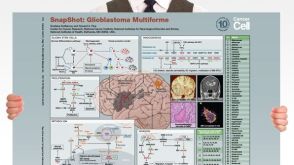 挂图SnapShot: Glioblastoma Multiforme Overview of the key concepts and mechanisms in glioblastoma multiforme biology
 沪公网安备31010102008431号